Improvement in Cysteine Production by Local Bacterial Isolates

Short Communications
Pakistan J. Zool., vol. 43 (4), pp. 805-808, 2011.
Improvement in Cysteine Production by Local Bacterial Isolates
Nazish Mazhar Ali,1 Farah Rauf Shakoori1* and A.R. Shakoori2
1Department of Zoology, GC University, Lahore
2School of Biological Sciences, University of the Punjab, Lahore
Abstract.-In the present study cysteine
producing bacteria from soil, water, milk, honey
and sewage were isolated with an aim of their
commercial exploitation. A total of 510 bacteria
were isolated, of which 51.3% produced some
amino acids viz. cysteine, methionine, lysine,
glutamic acid and valine. 1.76% of the total
isolates produced significant amount of
cysteine. The amino acid producing capability
of the bacteria was improved using different
types of fermentation media based on glucose,
urea, molasses, corn steep liquor, vitamins and
minerals. The composition of this medium
included glucose 40g, KH2PO4, 0.5g,
MgSO4.7H2O 0.3g and yeast extract 1.0g.
Bacterial strain MM5 produced 8.76g of
cysteine per litre in fermentation medium FM8.
The cost of this medium was Rs. 90.5/litre. The
cost of L-cysteine 10g is Rs. 2041 which is
quite costly as compared to the cost of this
medium. Other strains produced cysteine in
mostly range of 2g/litre. This medium was cost
effective and has potential for commercial
exploitation.
Key words:Amino acid producing bacteria,
microbial production of cysteine, commercial
production of cysteine.
A mino Acids are building blocks of the body, form antibodies to combat invading foreign bodies and act as source of energy (Abe and Takayma, 1972). Overproduction of amino acids by bacteria at commercial level is achieved by fermentation. Different wild and mutant strains of bacterial isolates have been used in microbial
_________________________
* Corresponding author: Telephone: 111-000-010, Ext. 457. 0030-9923/2011/0004-0805 $ 8.00/0
Copyright 2011 Zoological Society of Pakistan. fermentation of amino acids. Some wild strains of Corynebacterium glutamicum and Bacillus sp. are used for overproduction of different amino acids particularly lysine (Auger et al., 2002). In Pakistan, microbial production of amino acids has not been much exploited. These amino acids are imported from various countries including China, Malaysia, Spain and Germany. There is need to explore the ways to utilize natural resources available locally to produce cysteine at commercial scale. Government of Pakistan is spending 1800 dollars per metric tons annually for import of cysteine. Cysteine is commonly used in food industry as antioxidant in bread and fruit juices. Cysteine is also used for flavouring soft and fresh bread. This amino acid combines with different sugars and produces a seasoning with a pleasant meat flavor. Cooked meals and light snacks are prepared using cysteine for vegetarians. Cosmetic Industry also has great demand of cysteine. Cysteine is used in hair perming process. Currently cysteine is not synthesized at commercial level in Pakistan.
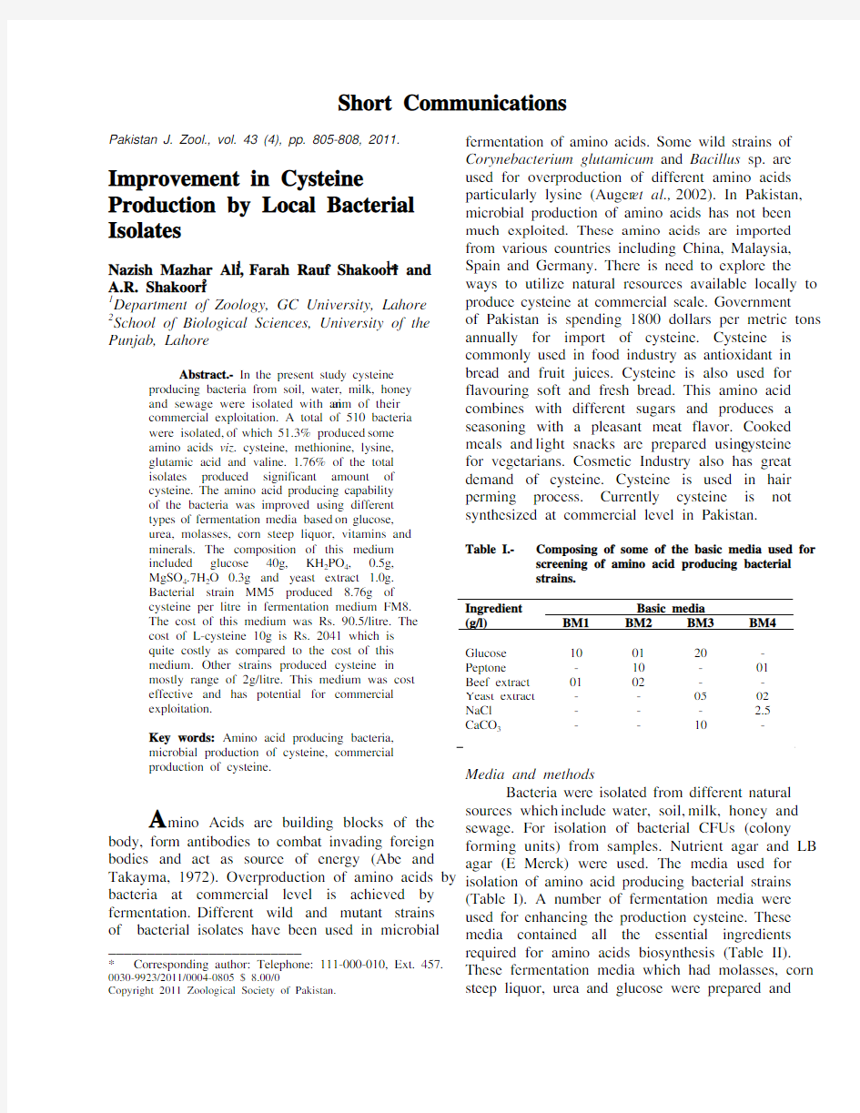
Table I.- Composing of some of the basic media used for screening of amino acid producing bacterial
strains.
Basic media
Ingredient
(g/l) BM1 BM2 BM3 BM4
Glucose 10 01 20 - Peptone - 10 - 01 Beef extract 01 02 - - Yeast extract - - 05 02 NaCl - - - 2.5 CaCO3- - 10 -
Media and methods
Bacteria were isolated from different natural sources which include water, soil, milk, honey and sewage. For isolation of bacterial CFUs (colony forming units) from samples. Nutrient agar and LB agar (E Merck) were used. The media used for isolation of amino acid producing bacterial strains (Table I). A number of fermentation media were used for enhancing the production cysteine. These media contained all the essential ingredients required for amino acids biosynthesis (Table II). These fermentation media which had molasses, corn steep liquor, urea and glucose were prepared and
SHORT COMMUNICATIONS 806
sterilized at 121oC and 15lb pressure for 15-20 minutes (Benson, 2002). The initial pH of most of the fermentation media was kept neutral, while others were slightly acidic in order to observe the tendency to bacterial strains to produce amino acids in media of different pH. In some fermentation media the pH was neutralized using calcium carbonate. The fermentation media FM1-7 were glucose based whereas FM8-12 were urea based whereas FM13-16, contained molasses. Few other fermentation media were also prepared containing vitamins e.g.biotin and other amino acids like leucine, threonine and other salts which could influence the production of amino acids by bacterial strains.
Table II.- Composition of some of the fermentation media for over production of cystein. FM8
supported production of 8.76g cystein per litre
of medium by isolate MM5.
Sr.No. Ingredient FM5 FM6 FM7 FM8
1 Glucose 27g 40g 30g 5.0g
2 Urea - - - 8.0g
3 K2SO420g - - -
4 KH2PO4 - 0.5g 2.0g 0.5g
5 MgSO4.7H2O 5.0g 0.3g - 0.2g
6 MnSO4.H2O 0.4g - - -
7 (NH4)2HPO420g - - -
8 NH4H2PO420g - - -
9 FeSo4.7H2O 0.2g - - -
10 Peptone - - - 2.0g
11 Meat extract - - - 2.0g
12 *CaCo3 - - 20g -
13 (NH4)2SO4.7H2O- - 10g -
14 Phenol red - - - 0.015g
15 Yeast extract 1.0g - -
16 MgCl2.4H2O - - 10mg -
17 FeSO4.4H2O 10mg -
18 Water 1000ml 1000ml 1000ml 1000ml
pH 7.0 7.0 7.2 7.0
*CaCO3 was added after sterilization of medium
Fermentation of amino acids
The screened bacterial strains were inoculated in 100ml of each fermentation medium in 250ml conical flasks. The flasks were incubated at 37±7oC in shaker at 125 rpm for maximum 96 hours. After every 24 h the 5ml sample was taken and cells were harvested. The harvest pH of each sample was recorded every time (Cheesebrough, 1993).
Analysis of amino acids
Five ml of fermented broth was taken out every 24 h centrifuged at 4000-5000rpm (2500xg) for 10min and supernatant used for analysis of amino acids. The supernatant was membrane filtered (0.45μm pore size) to make it cell free. The pellet was dried on filter paper at 70oC for 48 h and weighed to determine cell mass (Hassan et al., 2003). The amino acids were analysed both qualitatively and quantitatively. Paper chromatography was used for qualitative analysis of amino acids. Whereas acidic ninhydrin method (colorimetric method) was used (Chinard, 1952) for quantitative analysis. To 50μl of culture supernatant added 550μl of ninhydrin re agent in 5ml screw capped pyrex tubes. Known concentrations of standard solutions of amino acids were prepared in same way. The tubes were heated for one hour in 100oC water bath. After that tubes were cooled at room temperature and 1600μl of glacial acet ic acid was added in these tubes. Optical density was recorded at 365nm for cysteine. Standard curve was prepared by taking known concentration of cysteine. Selected amino acid producing strains were identified and characterized using different morphological and biochemical tests and determining nucleotide sequencing of 16S rRNA (ribotyping).
Table III.- Amino acids producing and non-amino acids producing bacterial strains isolated from
different sources.
Source
Total
no. of
bacterial
isolates
Producers
Non-
producers
Best
producers
Water 100 25 75 - Soil 250 189 61 05 Milk 50 05 45 - Honey 60 23 37 02 Sewage 50 20 30 02 Total 510 262 248 09
Results and discussion
From 510 bacterial isolates, 100 were isolated from water, 250 were isolated from soil, 50 from milk and 60 and 50 from honey and sewage. Out of a total of 510, overall 262 strains produced different amino acids. Of these only 9 strains, MM1-9 (Table V) produced cysteine in significant quantities. The
SHORT COMMUNICATIONS 807
overall percentage of producers was 51.37% and that of best producers was 1.76% (Tables III, IV). One bacterial strain was selected which was unable to produce amino acids and it was named as MM-ctrl. MM5 strain showed maximum production of
Table IV.- Amino acids producing bacterial strains
isolated from different sources during primary screening
Table V.- The amount of cysteine produced by different isolates after 96 hours of fermentation in different fermentation media.
Strain Fermentation medium Cysteine (g/l) Harvest pH
MM1 FM6 2.28 6.0 MM2 FM15 2.3 6.9 MM3 FM22 2.0 6.05 MM4 FM6 2.1 5.89 MM5 FM8 8.76 6.0 MM6 FM8 1.26 6.02 MM7 FM15 2.1 6.9 MM8 FM15 1.99 6.9 MM9 FM23 2.0 6.06
A m o u n t o f c y s t e i n e p r o d u c e d (g /l )
Time interval (hours)
Fig. 1. Amount of cysteine produced by MM5
cysteine among nine strains which is 8.76g/litre in FM8 medium (Fig. 1). The cost of FM8 medium was Rs. 90.5/- for one litre of medium. MM5 bacterial strain was isolated from sewage sample. MM5 strain was identified as Streptomyces sp. using biochemical tests and ribotyping technique (Figs. 2, 3). Amino acids are of great importance for metabolic activities in humans to ensure that body
O p t i
c a l
d
e n s i t y (600n m )
OPtimum pH
Fig. 2. Optimum pH of bacterial strain MM5
O P t i c a l d e n s i t y (600n m )
O ptimum Temperature (celsius)
Fig. 3. Optimum temperature of bacterial strain MM5.
Amino acids produced (per litre) Source
10-500 μg
501- 1000 μg 1.1- 50 mg 50.1- 100 mg 100.1- 999 mg 1g or
above
Water 25 - - - - - Soil 52 40 26 37 19 05 Milk 05 - - - - - Honey 30 15 05 05 03 02 Sewage 28 10 02 04 04 02
SHORT COMMUNICATIONS 808
performs its functions properly. The body excluding water comprises of seventy five percent amino acids. All neurotransmitters, except one, consist of amino acids and ninety five percent hormones are amino acids. L-cysteine, an important and well-known S-containing amino acid, has been widely used as a medical intermediate, and food or cosmetic additive. Traditionally, industrial L-cysteine production mainly depends on acid or alkaline hydrolysis of human or animal hairs (Yangjian et al., 2002). L-Cysteine is very important amino acid both biologically and commercially. Although most amino acids are commercially produced by fermentation, cysteine is mainly produced by hydrolysis of proteins. However, synthetic or biotechnological products have been available in the market. It was observed that using fermentation medium based on different natural ingredients, the cost can be minimized. All these bacterial strains are mesophiles and are locally isolated from soil, sewage and honey. Strain MM5 can be further improved for over-production of cysteine using further modified fermentation media or other biotechnological procedures. References
Abe, S. and Takayma, K.,1972. Amino acids producing micro-organisms, variety and classification. In: The microbial
production of amino acids, Kodanash, Tokyo, pp. 3-38. Auger, S., Yuen, W.H., Danchin, A. and Martin-Verstraete, I., 2002. Microbiology,148: 507–518.
Benson, H.J., 2002. Microbiological applications.8th ed. Mc-Graw Hill, New York.
Cheesebrough, M., 1993. Medical microbiology: Manual for tropical countries, vol. II. Microbiology ELBs,
University Press, Cambridge.
Chinard, F.P., 1952. J. biol. Chem.199: 91-95.
Hassan, B., Asghar, M., Nadeem, S. and Zubair, H., 2003. J.
Biotech., 1:18-29.
Khan, S.H., Rasool, G. and Nadeem, S., 2006. Pak. J.agric.
Sci., 43:
Theodora, T. and Bustard, T., 2005. Process Biochem.,40: 499-508.
Yangjian, L., Yingzi, Z., Jiang, W. and Yu, W., 2002. Acta microbiol. Sin., 42:395-399.
(Received 3 September 2010, revised 30 December 2010) Pakistan J. Zool., vol. 43(4), pp. 808-0, 2011.
RT-PCR Evaluation of Foot and Mouth Disease Serotype O in Saliva, Tracheal and Vesicular Samples of Goats in Punjab, Pakistan
Ali Saeed,1,2 Usman Waheed,3 Memoona Arshad,2 Qaiser Mahmood Khan, 2 Muhammad Ali1* and Muhammad Babar1
1Institute of Bio-Technology, Bahauddin Zakariya University, Multan, Pakistan
2National Institute for Biotechnology and Genetic Engineering (NIBGE), Faisalabad, Pakistan
3College of Veterinary and Animal Sciences, Jhang Sub-campus, University of Veterinary and Animal Sciences, Lahore, Pakistan
Abstract.- The aim of present study was
to evaluate the RT-PCR technique in the
original field samples from goat for the
diagnosis of foot and mouth disease (FMD)
within 24 hours. A total of 280 samples were
collected from thirty herds of goats in twelve
different outbreaks clinically suspected for
FMD near outbreak epicenters of other
livestock and evaluated by RT-PCR. with 171
positive results. Universal primer pair P1/P2
detected 30% and 1F/1R detected 61% from
total collected samples. AU(O)/AU(rev) and
AU(O)/PNK61 primer pairs confirmed O
serotype of FMD in 90.6% and 74.4%
respectively out of 43 representative samples,
whilst O-1C(ARS4)/ PNK61 O primer pairs
detected only 18.6% samples. The VP1gene
sequence analysis revealed O serotype of FMD
PanAsia1 lineage. The maximum samples can
be detected by 1F/1R, AU(O)/AU(rev) and
AU(O)/PNK61 primer pairs; whilst P1/P2,
AU(O)/AU(rev) and ARS4/NK61 can be a
better combination for the sequence analysis
from original field materials of goats.
Key words:FMDV (Foot-and-mouth disease
virus), PCR, field materials, goats
P anAsia 1 linage O serotype of foot-and-mouth disease (FMD) has replaced most of the ______________________________
* Corresponding author: alisam007@https://www.wendangku.net/doc/6e2529287.html,
SHORT COMMUNICATIONS 809
existing serotypes of FMD during previous years in Pakistan (Knowles et al., 2001; Saeed et al., 2009). Despite representing the majority of the world’s FMD susceptible livestock, sheep and goats have generally been neglected with regard to their epidemiological role in the spread of FMD (Patil et al., 2002).
The reverse transcription polymerase chain reaction (RT-PCR) has been shown to be a useful tool in the diagnosis of FMD in many previous studies, as a part of the viral genome can be detected within 24 hours in variety of samples with a wide range of primers targeting the different regions from the genome of FMD (Clavijo et al., 2003).
Foot-and-mouth disease was suspected on clinical observation e.g.high fever, lameness and rear mouth or foot lesions in number of goats during 2009 outbreaks in Pakistan. Many cases remained undiagnosed due to subclinical or similar clinical signs in many diseases of small ruminants. The study was designed to evaluate RT-PCR to detect FMD in clinically positive original field materials from goats. This study divulged importance and improvement of RT-PCR technique to detect FMD within 24 hours without virus isolations and in a variety of hosts especially in goats for fast, reliable and specific diagnosis of FMD virus directly from original field materials. Materials and methods
A total of 280 samples (saliva, tracheal and vesicular swabs) were collected from thirty herds of goats in twelve outbreaks clinically suspected for FMDV during 2009 in Sargodha, Faisalabad and Multan districts of Punjab, Pakistan. Saliva was collected by sterile ladies tampons, whilst the swab samples were collected with ear cleaning cotton sterile buds directly from trachea or site of vesicle into sterile falcon tube with 1.5 ml of transport medium (glycerol at equal amounts and 0.04 M phosphate buffer (pH 7.2), and the Penicillin (1000 IU/ml). Tissue samples were saved in transport medium and blood was collected in BD Vacutainer?. Finally, all suspected samples were transported to animal virology laboratory NIBGE. RT-PCR was performed by using the same primer pairs and protocol as described by Saeed et al. (2009). Results and discussion
FMD infected thirty herds of goats in twelve different outbreaks clinically suspected for foot-and-
mouth near outbreak epicenters of other livestock,
with 171 positive results. A number of goats were
also presented the clinical signs e.g.vesicular lesions in mouth, and lameness without any foot
lesion and off-feed due to high fever.
Universal primer pair P1/P2 from VP1 gene detected 84 (30%) out of 280 samples, whilst 1F/1R from 5' UTR region detected the 171 (61%) out of 280 original field materials. P1/P2 detected 42, 39
and 5 form saliva, mouth tracheal and mouth vesicle
swab samples respectively; whilst 1F/1R detected 61, 81 and 12 from saliva, mouth tracheal and mouth vesicle swab samples respectively (Table I). 1F/1R detected 87 more saliva, mouth tracheal and mouth vesicle swabs than P1/P2.
Similar to previous study by Saeed et al.
(2009), 1F/ 1R primer pair was found 31% superior
for primary diagnosis of FMDV all serotypes than universal primer set P1/P2 also in goats from original field materials. These finding also divulged the importance of virus isolation along with RT-PCR for complete detection of FMD in all suspected samples of clinically positive goats to monitor the carrier status or subclinical disease in these animals. Most of negative saliva and swab samples with RT-PCR were collected from the goats with no clinical lesion except fever.
The direct sequencing of VP1 gene PCR product of P1/P2 revealed that O serotype of FMD is still circulating in this region and this time appeared a little severe clinical disease in small ruminates also. O-1C (ARS4)/PNK61), AU(O)/ PNK61 and AU(O)/AU(rev) primer pairs were evaluated on 43 representative samples for the confirmation of FMD O serotype from original field material.
Genetic variation in Asian isolates and large
genome target made ARS4/NK61 less successful to detect the serotype O direct in original field materials but its importance cannot be neglected in molecular epidemiological work. FMD O serotype specific primer pairs AU(O)/AU(rev) and AU(O)/PNK61 can detect O serotype directly in clinically positive samples more efficiently (Reid et al., 2000; Knowles et al., 2001; Saeed et al., 2009).
SHORT COMMUNICATIONS
810
Table I.- PCR positive samples with different primer pairs found in total number of samples from goats during 2009.
Sample collection during 2009
Saliva, tracheal and vesicular swab samples processed Primer Pairs Total samples Positive samples
Saliva MTS MVS
Total samples processed with universal primers 280 171* 79 80 12 P1/P2 detected 280 84(30%) 42 39 5 1F/1R detected 280 171(61%) 68 81 12 Total samples processed with serotype specific primers 43 39* 14 15 10 O-1C (ARS4)/PNK61 detected 43 8 (18.6%) 3 4 1 AU(O)/AU(rev) detected 43 39 (90.6%) 14 15 10 AU(O)/PNK61 detected 43 32 (74.4%) 14 15 7 Primers (Saeed et al ., 2009)
MTS, Mouth tracheal swabs; MVS, Mouth vesicle swab
Bold (The main values), Non-bold (the subdivisions of the main values in both sides e.g. in rows and in columns)
Similarly, AU(O)/AU(rev) and AU(O)/ PNK61 primer pairs confirmed O serotype of FMD in 39 (90.6%) and 32 (74.5%), respectively out of 43 representative samples, while O-1C(ARS4)/ PNK61 O serotype-specific primer pairs detected O serotype of FMD only in 8 (18.6%) original field materials. Out of 39 serotype O positive samples saliva, mouth tracheal swabs and mouth vesicle swabs were 14, 15 and 10 respectively. The cloning and sequencing analysis of VP1 gene confirmed the serotype O of FMD in all samples (Table I). FMD in Asian countries due to uncontrolled movement of livestock is very difficult to control (Kitching, 1998). In sheep and goats, symptoms are frequently less severe and made the detection of the FMD very difficult (Knowles et al ., 2001). The outbreaks of FMD throughout the year are threatening for livestock trade within Pakistan and neighboring countries. Control of FMD in Pakistan requires proper strategy to diagnose it early both in large as well as small ruminants. FMD severe form of disease in the goats has been reported with high fatality rate in India (Uppal, 2003). Many FMD outbreaks in cattle in India are linked with the transmission of FMD virus from small ruminants. Hence, it is of paramount importance to diagnose this disease in early stages in small ruminants so that control measures can be adopted. FMD infected goats with severe clinical disease e.g. vesicular lesions in mouth, and lameness without any vesicular lesion on feet and
off-feed due to high fever etc in Sargodha, Faisalabad and Multan districts of Punjab during 2009. In this study, RT-PCR remained more successful to diagnose all twelve outbreaks but it detected only 61% original field samples collected from these suspected herds. Tracheal and mouth swabs collected with sterile ear cotton buds and saliva sample collected with sterile ladies tampons were better samples for RT-PCR analysis to detect FMD from original field samples. The mouth vesicle lesion appeared only in a few goats and mouth vesicle swab samples were not proved better samples for RT-PCR analysis direct from clinical sample. In conclusion maximum number of samples can be detected by 1F/1R, AU(O)/AU(rev) and AU(O)/PNK61 sets primer pairs; whilst P1/P2, AU(O)/AU(rev) and ARS4/NK61 are better combination for the sequence analysis and molecular epidemiological studies of FMD direct from original field material of goats.
Acknowledgements The authors thank extension teams for proper collection and transportation of samples.
References
Clavijo, A., Viera-Pereira, P.J. and Bergmann, I., 2003. Vet.
Res. Comm ., 27: 63–71.
Kitching, R.P., 1998. J. Comp. Pathol ., 118: 89-108.
Knowles, N.J., Samuel, A.R., Davies, P.R., Kitching, R.P. and
SHORT COMMUNICATIONS 811
Donaldson, A.I., 2001. Vet. Record, 148: 258-259. Patil, P.K., Bayry, J., Ramakrishna, C., Hugar, B., Misra, L.D., Prabhudas, K. and Natarajan, C., 2002. J. clin.
Microbiol., 40: 4367–4371.
Reid, S.M., Ferris, N.P., Hutchings, G.H., Samuel, A.R. and Knowles, N.J., 2000. J. Virol. Meth., 89: 167–176. Saeed, A., Khan, Q.M., Waheed, U., Arshad, M., Asif, M. and Farooq, M., 2009. Comp. Immunol. Microbiol. Infec.
Dis. doi:10.1016/j.
Uppal, P.K., 2003. Foot-and-mouth disease in small ruminants – An Issue of Concern (190 Appendix 29). Diagnostic
Research Laboratories, RWITC, 6 Arjun Marg, Pune
411 001, India.,190-194.
(https://www.wendangku.net/doc/6e2529287.html,/ag/againfo/commissions/docs/greece04/
App29.pdf)
(Received 24 August 2010, revised 25 October 2010) Pakistan J. Zool., vol. 43(4), pp. 811-814, 2011. Phenylthiocarbamide Tasting and its Implications – A Preliminary Study on Human Population Genetics in the Hazara Division of Pakistan
Faiza Rehman1, Arshid Pervez2*, Muhammad Awais2 and M.M. Shah1
1Department of Environmental Sciences, COMSATS Institute of Information Technology, Abbottabad, Postal Code 22060, Khyber Pakhtun Khwa Province, Pakistan.
2Life Sciences Services Center, Department of Environmental Sciences, COMSATS Institute of Information Technology, Abbottabad, Postal Code 22060, Khyber Pakhtun Khwa Province, Pakistan.
Abstract.- This study examined percentage of tasters and non tasters for
phenylthiocarbamide (PTC) in 325 subjects of
Hazara division in Khyber Pakhtun Khwa
Province of Pakistan. The percentage of tasters
was 87.7 and that of non-tasters 12.3.The sex,
age, food preferences (like vegetables, meat,
pulses, hot and very hot tea) indicated no
significant difference between tasters and non-
tasters. However, tasters showed affinity
towards salty food, whereas non tasters showed
easy acceptance to bitter medicines as compared
to the tasters.In addition higher percentage of
_____________________________
* Corresponding author pervez@https://www.wendangku.net/doc/6e2529287.html,.pk
super tasters was found among Swatis and
tasters among Tanolis.The genotype and gene
frequencies of tasting and non-tasting ability for
PTC indicated minute significant difference
between the parental and offspring generation.
Keywords: Phenylthiocarbamide (PTC), PTC
and race, PTC and food, PTC and genetics.
S ome human beings are taste blind to phenylthiocarbamide (PTC) and to 6-n-propylthiouracil (PROP). The human tongues have taste buds and fungiform taste papillae, the number of which is proportional to PTC/PROP taste (Fox, 1931). PTC/ PROP tasters have more taste pores than non-tasters, just as super tasters have anatomical differences in their fungiform papillae (Miller and Reedy, 1990).
Differences in the perception of taste and non-taste oral sensations result from a number of factors including, but not limited to, gender (Bartoshuk et al., 1994), age (Wise et al., 2007), ethnicity, salivary composition and flow rate (SFR), experience and environment (Yu and Pickering, 2008). However, the most important factor in the differences between individuals in perception of oral stimuli is genetic variation (Duffy, 2009). Any association between genetically mediated taste responsiveness to PTC and the acceptance of bitter phytochemicals might have implications for the design of dietary strategies for disease prevention (Looy and Weingarten, 1999).
Tasting is regarded as a fully developed genetic character. The two types may differ in their thresholds and it is possible to assign approximate probabilities of homozygosity to particular thresholds (Guo and Reed, 2001). A difference in the thresholds for PTC homozygous and heterozygous tasters could be established with certainty by testing people of known genotypes as inferred from their parents, siblings or children (Haris and Kalmus, 1949).
The main objective of this study was to determine the distribution and genetics of tasters, non tasters and super tasters in six different castes (three native and three migrated) from Hazara Division of Khyber Pakhtun Khwa, Pakistan. The Hardy-Weinburg equilibrium with respect to tasting
SHORT COMMUNICATIONS 812
ability in either generation separately and in both the generations combined was also tested.
Materials and methods
Fifty families from Hazara division, Khyber Pakhtun Khwa, Pakistan were tested for PTC tasting. Survey was conducted among the people both the males and females of 3-75 years of age of six major tribes living in the area. Subjects were excluded if they reported symptoms like cold, flu or any type of food allergy, whereas smoking was ignored. Subjects were made to place the impregnated filter paper (Drewnowski, 1997) on the tongue, moisten it to test for its bitterness. The results were recorded on the spot.
The nomenclature of PTC tasting used by Lim et al. (2008) was adopted according to which super tasters were able to perceive the bitterness of PROP at very low concentrations, moderate tasters perceived it at moderate concentrations, and non tasters were minimally or non-responsive even at high concentrations.
To determine relationship of tasting and non tasting ability with age, samples of 325 subjects were tested, and then phenotype frequencies of tasters and non tasters in these groups were compared.
The significance of relationship of tasting ability for PTC with gender, age, food preferences and caste was determined using chi-square test.
To test the Hardy-Weinburg demonstration that the genotype frequencies remain stable from one generation to the other, under certain conditions the general binomial theorem was used. Basic relationship when two alleles of a gene (Tt) are involved is shown as p+q= 1, where p is frequency of T allele (T for tasting ability), and q is frequency of t allele (t for non tasting ability). The expected genotypic or phenotypic frequencies in the next generation was summarized as (p+q)2= p2+ 2pq +q2=1, where p2is the fraction of the next generation expected to be homozygous TT, 2pq is the fraction expected to be heterozygous Tt and q2 is the fraction expected to be homozygous tt.
The phenotype frequencies for tasting and non-tasting in the samples of both the generations were observed. As we did not know the exact number of tasters having the genotype TT and Tt, we used non tasters having genotype tt of both generations as a tool for calculation of the gene frequencies, using the binomial theorem of Hardy-Weinberg equation. The gene frequencies of T and t in both generations were expressed by p and q for T and t, respectively, in the two generations.
The allelic frequencies in the two generations was compared by using z-test:
Z = P1-P2
√pq(1/n1 + 1/n2)
Results and discussion
Studies on tasting ability for PTC were often limited to college students, mostly women and to relatively small subject samples (Kobayashi and Kennedy, 2006; Lim et al., 2008) but the present study was conducted both in males and females of general population. In this study the comparisons between tasters, non tasters and super tasters with gender indicated no significant differences providing evidence that PTC tasting is not dependent on gender (Table I), whereas according to Drewnowski (2001), the percentage of non-tasters for PROP increased with age in both men and women.
The data in Table I shows that tasting ability was not affected by age or did not change with increasing age.
The study comparison of tasters, non-tasters and food preferences like vegetables, meat pulses, hot or very hot tea indicated no significant difference while 82.5% of tasters and 65% of non-tasters preferred sweet food. Thus presence of tasting ability for PTC had greater affinity towards sweet food as compared to the non-tasters. Similarly 50.9% of tasters and 72.5% of non-tasters preferred salty food, indicating greater affinity of non-tasters towards salty food. 67% of non-tasters felt no difficulty during intake of bitter medicine showing their easy acceptance to bitter medicines as compared to the tasters (Table II). Also in a comparison between tasters, and super tasters 83.05% of super tasters and 46.90% of tasters felt difficulty during intake of bitter medicine.
The comparison of non tasters, super tasters indicated no significant difference. However 65% non tasters and 84.74% super tasters showed vivid
SHORT COMMUNICATIONS 813
Table I.- Correlation between tasters, non tasters, super tasters and gender and age.
Tasters Non-tasters Tasters Super-tasters Non-Tasters Super-tasters Gender
Male
Observed 133 22 110 29 22 29 Expected 135.92 19.1 114.23 29.82 20.61 30.39 Female
Observed 152 18 116 30 18 30 Expected 149.1 20.92 119.98 31.32 19.39 28.60
χ2 0.96χ2 = 0.308χ2 = 0.308 (df= 1, α = 0.05) Age
3-15 yrs
Observed 77 9 64 13 9 13 Expected 75.41 10.58 61.06 15.94 8.8 13.2
16-30 yrs
Observed 104 9 78 26 9 26 Expected 99.09 13.91 82.47 21.53 14 21
31-45 yrs
Observed 56 10 43 13 10 13 Expected 57.88 8.12 44.41 11.49 9.2 13.8
46-60 yrs
Observed 45 12 38 8 12 8 Expected 49.98 7.01 35.68 9.31 8 12
61-75 yrs
Observed 3 0 3 0 0 0 Expected 2.63 0.37 2.38 6.62
χ2 = 8.47χ2 = 2.55χ2 = 6.327 (df= 4, α = 0.05) Table II.- Correlation between tasters, non-tasters, super taster and food preferences and castes.
Tasters Non-
tasters χ2Tasters Super-
tasters
χ2Non-
Tasters
Super-
tasters
χ2
Food items
Vegetables 165/285 24/40 0.057 127/226 38/59 1.28 24/40 38/59 0.19 Pulses 223/285 26/40 3.43 178/226 45/59 0.147 26/40 38/59 0.003 Meat 195/285 32/40 2.23 156/226 39/59 0.18 32/40 39/59 2.19 Sweet food 235/285 26/40 6.67 185/226 50/59 0.24 26/40 50/59 5.08 Salty food 145/285 29/40 6.57 103/226 42/59 12.28 29/40 42/59 0.018 Hot tea 201/285 29/40 6.57 162/226 39/59 0.69 29/40 39/59 0.437 Very hot tea 84/285 11/40 0.07 64/226 20/59 24.52 11/40 20/59 0.437 Difficulty in bitter
medicine intake
155/285 13/40 6.7 106/226 49/59 24.52 13/40 49/59 25.93 No difficulty in bitter
medicine intake
130/285 27/40 6.7 120/226 10/59 24.52 27/40 10/59 25.93
Castes
Awan 86/285 16/40 1.57 65/226 21/59 1.02 16/40 21/59 0.189 Swati 73/285 10/40 0.007 51/226 22/59 5.32 10/40 22/59 1.5 Tanoli 44/285 6/40 0.004 40/226 4/59 4.26 6/40 4/59 1.71 Rajput 43/285 7/40 0.16 37/226 6/59 1.4 7/40 6/59 0.93 Bhatti 16/285 1/40 12.68 14/226 2/59 0.69 1/40 2/59 0.05 Abbasi 23/285 0/40 3.37 19/226 4/59 0.16 0/40 4/59 2.8
SHORT COMMUNICATIONS 814
affinity towards sweet food. Moreover, 67.5% of non tasters and 16.94% of super tasters felt no difficulty in intake of bitter medicines. This means, among the tasters the subgroup of super tasters showed more rejection to bitter medicines as compared to tasters (Table II).
The overall percentage of tasters in Hazara division was 87.70% and that of non-tasters 12.30%. The frequency of PTC non-tasters was estimated to be 10% in China (Guo and Reed, 2001). Among the Europeans, taster/non-taster status was about evenly divided, but among Asians, 70% were tasters (Miller and Reedy, 1990). The proportion of PTC tasters in the US population was estimated as 70% (Bartoshuk et al., 1994). The known variation in the number of non-tasters across race almost assured that when super-taster categorization was available, they vary across race (Bartoshuk et al., 1994). Early studies suggested that the ability to taste PROP was more common among Asians and Africans than among Caucasians. According to the present study with six different castes, Swatis, Bhattis and Tanolis had different percentages of tasters and super tasters suggesting that the ability to taste has some relation to race or caste in an observed population (Table II). Also the percentage of a subgroup of tasters i.e. super tasters vary from caste to caste. Significant difference existed between tasters and super tasters of Swati and Tanoli castes. Among Swatis 22.6% were tasters and 6.7% were super tasters.
The present data includes two successive generations, 130 parents 110 tasters, 20 non-tasters and 195 off springs (175 tasters, 20 non-tasters).
There existed no significant difference. The calculated value was less than the tabulated value. Thus the conclusion that the allelic frequencies in the two generations were comparable could not be drawn with certainty.
As it is obvious that different genotypes produced through random mating depends upon the gene frequencies of the parental generations. Therefore, by using frequencies of the allelic frequencies of parental generation we calculated and compared the expected phenotypic frequencies of the offspring generation (Table III).
In the case of Hardy Weinburg equilibrium existing of minute significant difference in comparison of observed phenotypic frequencies in the offspring generation with those expected from parental gene frequencies assuring random mating among them, created a chaotic situation (Table IV). Thus no conclusion could be drawn with certainty in this regard.
Table III.- Calculated gene frequencies of T and t in the two generation.
Generation p (T) q (t) Parents 0.608 0.392 Offsprings 0.68 0.32
Table IV.- Comparison of expected and observed phenotypic off spring frequencies . Generation Taster Non-taster Offspring
Observed 175 20 Expected 165 30
χ2 = 3.941 (df = 1; α = 0.05) [Minute significant difference] References
Adam, D. and Agnes, L.Y., 2001. Chem. Senses,26: 41-47. Bartoshuk, L.M., Duffy, V.B. and Miller, J.I., 1994. Physiol.
Behav., 56: 1165-1171.
Drewnowski, A., 1997. Ann Rev Nutr, 71: 237-253.
Duffy, V.B., Hayes, J.E., Bartoshuk, L.M. and Snyder, D.J., 2009. Encyclop. Neurosci., 5: 881-886.
Fox, A.L., 1931. Sci. News, 9: 249.
Guo, S.W. and Reed, D.R., 2001. Annl. Hum. Biol,, 281: 11-42. Haris, H. and Kalmus, H., 1949. Ann. Engen.,15: 24-34. Kaminski, L.C., Handerson, S.A. and Drewnowski, A., 2000.
Physiol. Behav., 68: 691-697.
Kobayashi, C. and Kennedy, L.M., 2006. Chem. Senses, 31: 301-306.
Lim, J., Urban, L. and Green, B.G., 2008. Chem. Senses,33: 493-501.
Looy, H. and Weingarten, H.P., 1999. Physiol. Behav., 52: 75-
82.
Miller, I.J. and Reedy, F.E., 1990. Physiol. Behav., 47:1213-1219.
Pickering, G.P., 2008. Am. J. Enol. Vitic, 59: 146-152.
Wise, P.M., Hansen, J.L., Reed, D.R. and Breslin, P.A.S., 2007.
Chem Senses, 32: 749-754.
Yu, P. and Bartoshuk, L.M., 1993. Fd. Qual. Pref., 4: 21-32.
(Received 3 August 2010, revised 29 November 2010)
SHORT COMMUNICATIONS 815
Pakistan J. Zool., vol. 43 (4), pp. 815-818, 2011.
Prevalence of Multiple Drug Resistant Staphylococcus aureus in Different Hospitals of Lahore
Numan Javed, Nadeem Afzal*, Romeeza Tahir, Afia Abbas and Khursheed Javeed
Department of Immunology, University of Health Sciences, Lahore, Pakistan
Abstract.- Staphylococcus aureus is a
multidrug resistant pathogen that causes a wide
range of infections in human beings. In order to
control the increase in infection rate, it is
necessary to screen the samples adequately.
Present study was conducted to find out the
biochemical characteristics and methicillin
resistance of S. aureus isolates from various
hospitals of Lahore, Pakistan. One hundred
bacterial isolates were collected from various
public and private sector hospitals. Fifty isolates
were declared as methicillin resistant S. aureus
(MRSA) while others as methicillin sensitive S.
aureus(MSSA) by performing Kirby-Bauer
disc diffusion method. Biochemical properties
were confirmed by Gram staining, mannitol
fermentation, catalase and DNase production.
Methicillin resistance was checked by chrome
agar for MRSA. In MRSA group 6 (12%)
isolates were not MRSA and 4 (10%) isolates
were coagulase negative staphylococci. While
in MSSA group 25 (28%) isolates were not S.
aureus. Probably such inappropriate screening
of organisms and unnecessary antibiotic
prescription are the risk factors for increase in
antibiotic resistance.
Key words:MRSA, MSSA, antibiotic
resistance
S taphylococcus aureus (S. aureus) are Gram positive cocci that normally reside in squamous epithelium of nasopharynx and vagina of approximately 5% females (Levinson, 2008). It causes a wide range of opportunistic infections e.g. abscesses, folliculitis, cellulitis, impetigo, central nervous system infections, urinary tract infections, chronic lung infections associated with cystic _________________________________
* Corresponding author: immunology@https://www.wendangku.net/doc/6e2529287.html,.pk fibrosis, infective endocarditis, septic arthritis, osteomyelitis, food poisoning, pneumonia, septicemia, surgical wound infections, scalded skin syndrome, toxic shock syndrome and Kawasaki’s syndrome (Loir et al., 2003; Smeltzer and Gillaspy, 2000; Gill et al., 2005).
Penicillin was developed in early 1940s to eradicate S. aureus infections but due to genomic plasticity the organism became resistant to this antibiotic (Lowy, 2003, Jeong et al., 2007). Thereafter, methicillin was developed in 1960 to treat penicillin resistant organisms but within a year evolution of methicillin resistant S.aureus (MRSA) was reported in England (Grundmann et al., 2006). Resistance against methicillin is encoded by mecA gene which is located on staphylococcal chromosomal cassette (SCC mec). SCC mec is made up of two segments; mec gene complex (A-E) which encodes antibiotic resistance and ccr gene complex that helps in excision and integration of whole genetic vector. The mecA gene encodes for altered penicillin binding protein-2a (PBP-2a) instead of normal penicillin binding proteins (PBPs), and it is suggested that altered PBPs has reduced affinity for methicillin (Kuroda et al., 2001; Ito et al., 2001, 2004). So far seven major types and variants of SCC mec elements have been reported in different parts of the world and each one of these is capable of encoding resistance to multiple antibiotics (Hanssen and Sollid, 2006; Shore et al., 2005).
The prevalence of MRSA varies among different geographic regions. In Asian countries such as Japan, Korea and China the incidence of MRSA has reached more than 70% (Ko et al., 2005). Prevalence of MRSA has dramatically increased in Pakistan over the past two decades as compared to other parts of the world and it is reported that in Pakistan the prevalence of MRSA is about 32%. In 2004, incidence of MRSA in different hospitals of Lahore was reported as: Sheikh Zayed Hospital (20%), Services Hospital (21.6%), Lahore General Hospital (32.6%) and Mayo Hospital (38.5%). As a consequence, antibiotic resistance is reaching at an alarming level because treatment options are becoming gradually limited (Bukhari et al., 2004; Perwaiz et al., 2007).
In order to control the rate of infection, it is necessary to precisely screen the pathogenic
SHORT COMMUNICATIONS 816
organisms in the shortest span of time. Depending upon the screening results, isolation procedures can be adopted and antibiotic regime can be prescribed. Goal of the present study was to check the biochemical characteristics and antibiotic sensitivity of S. aureus isolates by chrom agar that are already characterized that are already identified by various hospitals of Lahore.
Materials and methods
It was a cross sectional and descriptive study that was completed in one year. One hundred bacterial isolates were collected from different public and private sector hospitals of Lahore. A well defined and single colony of S. aureus was transferred from nutrient agar plate to brain heart infusion broth (BHI, Oxoid, UK) and it was incubated overnight at 37 °C. These stains were maintained in brain heart infusion containing 25% glycerol (Sigma, St. Louis) at -20° C. In our laboratory, all the bacterial strains were confirmed by Gram staining, mannitol fermentation, catalase and DNase production. MRSA and MSSA strains were screened by using chrom agar (Beckton, Dickinson BBL) for MRSA (Cheesbrough, 2004). Results and discussion
There were fifty isolates of MRSA which comprised of 41 (82%) from one public sector hospital, 03 (06%) from another public sector hospital while 06 (12%) isolates were from a private sector hospital. The other fifty isolates were MSSA. Among MSSA group, 22 (44%) isolates were collected from one public sector hospital, 04 (8%) from another public sector hospital and 24 (48%) from a private hospital of Lahore. One hundred confirmed bacterial isolates of S.aureus were collected from different hospitals of Lahore which included fifty MRSA and fifty MSSA isolates each.
On screening MRSA isolates from one public sector hospital, it was found that 06 (12%) isolates were not MRSA and another 04 (10%) isolates were not even S. aureus, rather these 04 isolates were coagulase negative staphylococci. However, all MRSA isolates from second public sector hospital and private sector hospital were MRSA (Fig. 1A).
Among MSSA group, on screening these isolates, it was found that 05 (22%) from one public sector hospital, 01 (25%) from another public sector hospital and 09 (37%) from private sector hospital were not S. aureus (Fig. 1B). Present study showed that 4 (9%) isolates declared as MRSA were actually found to be coagulase negative
staphylococci in our laboratory.
Fig. 1. Occurrence of MRSA (A) and MSSA (B) in three different hospitals of
Lahore.
SHORT COMMUNICATIONS 817
Prevalence of MRSA in different regions of Pakistan varies from 5% to 42%. Infections caused by resistant pathogens usually resulted in prolonged illness, financial burden and higher risk of mortality (Cosgrove et al., 2005). Drew et al. (1972) stated that in routine practice many organisms identified as MRSA were actually coagulase negative S. epidermidis and infect, they were misidentified in other laboratories. The reason might be that, S. epidermidis is normally present on skin and whenever skin samples are not collected under sterile conditions, there are chances of contamination with this organism. Evolutionary data suggested that mecA gene might have transferred to S. aureus from S. epidermidis in vivo (Lencastre et al., 2007). Therefore in order to avoid catastrophic consequences of multi resistance, it is highly suggested to properly deal with these organisms. It was found in the current study that 6 (13%) isolates were MSSA whereas they were reported as MRSA. Similarly 5 (19%) isolates were MRSA but reported as MSSA. Chrom agar MRSA is a differential and selective medium for detection of MRSA. It is incorporated with chromogenic substrate and cefoxitin to inhibit the growth of sensitive strains. MRSA grows in the presence of cefoxitin and produces mauve coloured colonies (https://www.wendangku.net/doc/6e2529287.html,/ds/technicalCenter/clsi/clsi-chromagarmrsa.pdf). In order to overcome the burden of resistant pathogens, it is necessary to precisely screen them by rapid, specific and sensitive methods for therapeutic purposes (Kunori et al., 2002; Stoakes et al., 2006). Inappropriate screening of methicillin resistance leads to an inaccurate diagnosis and incorrect prescription of antibiotics. Currently, misidentified MRSA which could be actually MSSA are being treated with vancomycin that is an ultimate choice of antibiotics rather being treated with methicillin. Further, it is documented that in the countries where the rate of MRSA infection is high, there is a tradition to start the treatment prophylactically with vancomycin whenever there are some signs of infection. It should be remembered that vancomycin is the drug of last resort against MRSA and it is expensive and it also eliminates normal flora that may lead to opportunistic infections (Enright, 2003).
Resistance against antibiotics is a natural evolutionary phenomenon but this process can be accelerated by human practices such as inappropriate screening of infecting organisms and unnecessary antibiotic prescription. Therefore antibiotic use must be confined to conditions where it is specifically indicated. A decrease in unnecessary antibiotic prescription can effectively reduce the rate of antibiotic resistant organisms (Mirza, 2007; Chinedum, 2005). From the study it can be concluded that misdiagnosis and false reporting of S. aureus as coagulase negative staphylococci and incorrect identification of resistant strains are serious threats for increase in antibiotic resistance.
Acknowledgements
We thank University of Health Sciences Lahore for providing funds for this research project. We would also like to express our gratitude to all the patients whose samples have been used to carry out the study.
References
Bukhari, M.H., Iqbal, A., Khatoon, N., Iqbal, N., Naeem, S. and Qureshi, G.R., 2004. Pak. J. med. Sci., 20:229-233. Cheesbrough, M., 2004. District laboratory practice in tropical countries. Cambridge University Press UK, pp. 157-59. Chinedum, I.E., 2005. Afr. J. Biotech., 4:1606-1611. Cosgrove, S.E., Qi, Y., Kaye, K.S., Harbarth, S., Karchmerr,
A.W. and Carmeli, Y., 2005. Infect Contr. Hosp
Epidemiol., 26:166-174.
Drew, W.L., Barry, A.L., O’Toole, R. and Sherris, J.C., 1972.
Appl. Microbiol., 24:240-247.
Enright, M.C., 2003. Curr. Opin. Pharmacol., 3:474-479. Gill, S.R., Fouts, D.E., Archer, G.L., Mongodin, E.F., DeBoy, R.T. and Ravell, J., 2005. J. Bact., 187:2426-38. Grundmann, H., Aires de-Sousa, M., Boyce, J. and Tiemersma,
E., 2006. Lancet, 368:874-885.
Hanssen, A.N. and Sollid, J.U., 2006. Immunol. Med.
Microbiol., 46:08-20.
Ito, T., Katayama, Y., Asada, K., Mori, N., Tsutsumimoto, K.
and Tiensasitom, C., 2001. Anti- Microbiol.
Chemother., 45:1323-1336.
Ito, T., Ma, X.X., Takeuchi, F., Okuma, K., Yuzawa, H. and Hiramatsu, K., 2004. Antimicrob. Agents Chemother.,
48:2637-2651.
Jeong, H.Y., Lee, J.E., Choi, B.Y., Seo, K.W., Park, S.H. and Kim, Y.L., 2007. Microb. Drug Resist., 13:178-185. Ko, K.S., Le, J.Y., Suh, J.Y., Oh, W.S., Peck, K.R. and Lee, N.Y., 2005. J. clin. Microbiol., 43: 421-426.
SHORT COMMUNICATIONS 818
Kunori, T., Cookson, B., Roberts, J.A., Stone, S. and Kibbler,
C., 2002. J. Hosp. Infect., 51:189-200.
Kuroda, M., Ohta, T., Uchiyama, I., Baba, T., Yuzawa, H. and Kobayashi, I., 2001. Lancet, 357:1225-1240. Lencastre, H., Oliveria, D. and Tomasz, A., 2007. Curr. Opin.
Microbiol., 10:428-435.
Levinson, W., 2008. Review of medical microbiology and immunology, 10th ed. McGraw Hill; New York. pp. 106-
118.
Loir, Y.L., Baron, F. and Gautier, M., 2003. Genet. mol. Res., 2:63-76.
Lowy, F.D., 2003. J. clin. Invest., 111:1265-1273.
Mirza, S.H., 2007. Infect. Dis. J.,19: 75-79.
Perwaiz, S., Barakzi, Q., Farooqi, B.J., Khursheed, N. and Sabir, N., 2007. J. Pakistan med. Assoc., 57: 02-04. Shore, A., Rossney, A.S., Keane, C.T., Enright, M.C. and Coleman, D.C., 2005. Antimicrob. Agents Chemother.,
49:2070-2083.
Smeltzer, M.S. and Gillaspy, A.F., 2000. Poult. Sci., 79:1042-1049.
Stoakes, L., Reyes, R., Daniel, J., Lennox, G., John, M.A. and Lannigan, R., 2006. J. clin. Microbiol., 44:637-639.
(Received 11 January 2010, revised 11 May 2010) Pakistan J. Zool. vol. 43 (4), pp. 818-821, 2011.
A Complicated Case of Septic Orchitis and Posthitis in a Race Horse
M. A. Khan*, M. M. Ali and S. Azeem Department of Clinical Medicine and Surgery, University of Veterinary and Animal Sciences Lahore, Pakistan
Abstract.- A ten year old bay stallion was presented with a history of scrotal swelling
along with posthitis and difficult micturation
(Stanguria). Palpation of the external genitalia
revealed variable consistency of various areas
of the affected organs. The animal was cast and
examined for scrotal herniation but no evidence
for it was found. The surgical exploration of the
lesions revealed septic orchitis. After surgery
stallion was treated with systemic antibiotic and
anti-inflammatory drugs and the animal
recovered completely within two weeks.
Key words: Stallion, scrotal swelling, scrotal
hernia, pus, castration.
__________________________
* Corresponding author: vetdrarif@https://www.wendangku.net/doc/6e2529287.html,
M ale genital organs of equids may encounter a number of abnormalities such as neoplastic growths, scrotal haematoma, abscessation in scrotum, scrotal hernia, orchitis, balanitis, and posthitis. The infection may extend from a wound or spread from hematogenous route. Furthermore, extension of infection from the accessory sex glands etc. has also been incriminated for genital disorders (Kasaback et al., 1999; Johnson, 1986). Most often than not, these anomalies are caused by trauma and are evident by swelling and enlargement of the male genital organs (Meadows and William, 1995). We report here a complicated case of septic orchitis and posthitis in a stallion (Vaughan, 1986).
Materials and methods
A ten year old bay stallion was brought to the Indoor Hospital, Surgery Section, University of Veterinary and Animal Sciences, Lahore, Pakistan for an evaluation of a profuse swelling on the scrotum extending up to ?th of the prepuce (Fig. 1A). The swelling was of approximately 10 days duration, however, the genital organs were not routinely examined and the lesions may have been of longer duration. Physical examination and palpation of the affected organs revealed variable consistency of different areas. The animal was treated systemically with prednisolone 375 mg and a combination of procaine penicillin 10000 I.U./kg, Benzyl pencillin 10000 I.U./kg, and streptomycin 11 mg/kg. However, medical treatment failed to give satisfactory response. The animal was cast under light sedation with detomidine at a dose rate of 40 μg/kg body weight intravenously and examined for scrotal hernia but no visceral contents were palpable. Needle aspiration revealed malodorous purulent discharge (Belknap et al., 1988). No ultrasonographic evaluation (Morresey, 2007) could be carried out due to non-availability of ultrasonography. It was decided to perform open castration (Meadows and William, 1995). The vital signs of the stallion were recorded on the day of operation and for five days postoperatively (Table I).
The present case presents a picture typical of septic orchitis (Belknap et al., 1988). Clinico-pathological abnormalities were indicative of internal infection that may have led to chronic
SHORT COMMUNICATIONS 819
A
B
C
Fig. 1. Surgical procedure for septic orchitis and posthitis in a race horse; A, swelling on scrotum and sheath before operation; B, Pus and serosanguinous fluid during operation; C, Surgical site plugged with gauze of tincture iodine.inflammatory/infectious process (Aleman et al., 1996). Thrombocytopenia and elevated total serum proteins were indicative of infection and suppurative disease and increased level of urea and blood urea nitrogen (Table II) revealed reduced renal blood flow (Benjamin, 1985) which may be a sequel of trauma.
Table I.- Vital signs of the animal.
Days Temperature
(°F)
Pulse
(beats per
minute)
Respiration
(breaths per
minute)
1 101.83614
2 101.23912
3 101.44014
4 101.03611
5 101.23813
Table II.- Hematological and serum biochemical profile of the patient.
Parameters Normal value Hematology
RBC (x1012/l) 9.12 (7-13)
WBC (x109 /l) 7.71 (7-14) Lymphocytes (%) 16 (25-70)
Monocytes (%) 6.5 (0.5-7) Granulocytes (%) 77.4 (30.5-79)
PCV (%) 31. 3(32-55) Hemoglobin (g/dl) 11 (10-18)
Platelets (109/l) 80 (200-400)
Serum biochemistry
Serum SGOT (μ/l)281 (79±11)
Serum SGPT (μ/l) 10 (11±1)
Serum urea (mg/dl) 52 (7-27)
Blood urea nitrogen (mg/dl) 24 (3.2-3.11)
Serum cholesterol (mg/dl) 46 (75-150)
Total serum protein (g/dl) 10 (6.0-7.9)
Serum creatinine (mg/dl) 1.14 (1.3-1.67) Reference values are shown in parenthesis. Recorded parameters that fall outside the reference range are shown in bold.PCV, packed cell volume; RBC, red blood cells; SGOT, serum glutamic oxaloacetic transaminase; SGPT, serum glutamic pyruvic transaminase; WBC, white blood cells.
The animal was kept fast 24 hours before surgery. Xylazine hydrochloride 0.5mg/kg was administered as a pre-anesthetic and chloral hydrate 1gm/10kg as a general anesthetic, intravenously. Prophylactic antitetnus serum was administered subcutaneously. Open castration was performed as follows; the horse was cast on left lateral
SHORT COMMUNICATIONS 820
recumbency. Before starting the operation, the scrotum and sheath were thoroughly cleaned with soap and water, followed by application of tincture of iodine as an antiseptic. A bold incision was made on the scrotal skin about ? inch lateral to the median raphae. An exposure of the lesion revealed necrosis of testes and dissolution of the median septum between the testes. Extension of the incision led to oozing of malodorous pus along with serosanguinous fluid (Fig. 1B). The vascular and avascular portions of spermatic cord of both the testes were crushed and cut with the help of an emasculator through the same window. After irrigating the scrotal rent with Tincture of iodine and removing all debris and dead necrotic material, it was packed with gauze impregnated with tincture of iodine. Stay sutures were applied on the incision to keep the gauze in place (Fig. 1C). The animal recovered from anesthesia in about 20 minutes after the surgery. A combination of chloramphenicol 120 gm, tetracycline 80 gm, sulphamethoxypridazine 100 gm and prednisolone 375 mg was administered post surgically via intramuscular route. The same systemic treatment was repeated for the following five days. Gauze was removed after one day of surgery and the surgical wound was treated as an open wound. A marked decrease in swelling was observed the following day. The animal’s owner was advised daily irrigation of the surgical site with clean fresh water for half an hour morning and evening.
Results and discussion
It is difficult to diagnose and manage the cases of enlarged scrotum in the stallion (Morresey, 2007). The scrotal swelling may result due to hydrocele, pyocele, septic orchitis, hematoma, neoplasia, testicular torsion, habronemiais, inguinal and scrotal hernia (Meadows and William, 1995; Morresey, 2007; Roberts, 1986). In the present case, orchitis was the result of trauma although no history of trauma was provided. In horses, trauma may result in septic orchitis and periorchitis but occasionally it can be of hematogenous origin (Kasaback et al.,1999; Belknap et al., 1988). Trauma is a common cause of scrotal swelling in the stallion. It may cause scrotal contusion and testicular hematoma (Morresey, 2007). Trauma can also cause intense inflammatory edema of both penis and prepuce (Vaughan, 1986). Orchitis is characterized by marked inflammation and abscessation in the vaginal sac. Moreover, diffuse edematous swelling may also be formed in the vicinity (O’Conner, 2005); this finding is consistent with the present case. Purulent orchitis in cattle and dogs, however, is usually associated with brucellosis (Hopkins, 1997; Johnson, 1986). Purulent orchitis is uncommon in Alpacas (Pascale et al., 2000).
The diagnosis of such cases is based on history, physical examination, palpation of testes and scrotum, aspiration cytology, semen collection and its evaluation. The diagnosis can be established by direct palpation of the scrotum (Morresey, 2007). Fluctuation on palpation is not a reliable diagnostic feature in such cases. It is necessary to aspirate the contents of the swelling with the help of a needle to differentiate between seroma, hematoma and abscess. A surgical incision is made on the scrotum for drainage (Vaughan, 1986). Needle aspiration and surgical incision on scrotum for drainage revealed septic orchitis. Castration is often the sole remedy for these conditions (Meadows and William, 1995; O’Conner, 2005).
Depending on the etiology and severity of enlargement, irreversible injury may result to the reproductive system and adjacent structures of a stallion. Permanent damage to the reproductive system may result as a sequel to scrotal enlargement depending on the cause and severity of the swelling. Scrotal swellings although appearing clinically similar have myriad etiologies.
Initially scrotal swelling was thought of a scrotal hernia, hydrocele, pyocele, septic orchitis, hematoma, neoplasia, testicular torsion but on surgical exploration of scrotum, purulent orchitis was revealed. Thrombocytopenia and increased level of total serum protein suggested infectious septic process (Benjamin, 1985).Increased level of urea and blood urea nitrogen may have been the result of urinary retention. Inflammation subsided after performing castration in the following days. The case was monitored postoperatively for 2 weeks; the animal recovered uneventfully and was sent back to the track. It is concluded that such cases should be reported to the clinic at an early stage for the efficient management and treatment.
SHORT COMMUNICATIONS 821
References
Aleman, M., Spier, S.J., Wilson, W.D. and Doherr, M., 1996. J.
Am. Vet. med. Assoc., 209: 804–809.
Belknap, J., Arden, W. and Yamini, B., 1988. J. Am. Vet. med.
Assoc., 192: 363-364.
Benjamin, M.M., 1985. Outline of veterinary clinical pathology, 3rd ed. Kalyani Publishers, Ludhiana, India.
pp.113-170.
Hopkins, F.M., 1997. Diseases of the reproductive system of the bull. In: Current therapy in large animal
theriogenology(ed. R.S. Youngquist). WB Saunders,
Philadelphia: USA. pp. 237-239.
Johnson, C.A., 1986. Disorders of the canine testicles and epididymides. In: Current therapy in theriogenology 2
(ed. D.A. Morrow), WB Saunders. Philadelphia. pp.
551-552.
Kasaback, C.M., Rashmir-Raven, A. M. and Black, S.S., 1999.
J. Am. Med. Assoc., 215: 787-789.
Meadows, A.W. and William, B.L., 1995. Advanced equine reproduction course. Available at URL:
http://www.horse-
https://www.wendangku.net/doc/6e2529287.html,/ARTICLES/PDFs/ScrotalSwelling.pdf.
Accessed on Jan., 09, 2011.
Morresey, P.R., 2007. Clin. techniq. equine Pract., 6: 265-270. O’Conner, J.J., 2005. Dollar’s veterinary surgery, 4th edn., CBS publishers and Distributors, New Delhi, India. pp. 745-
746.
Pascale, A., Tamara, M.S., Christiane, V.L., Ahmed, T. and George, M.B., 2000. Can. Vet. J., 41:704-706. Roberts, S.J., 1986. Veterinary obstetrics and genital diseases.
Woodstock, VT. USA. pp. 752-893.
Vaughan, J.T., 1986. Surgery of the male reproductive system.
In: Current therapy in theriogenology 2 ed. (ed. D.A.
Morrow), WB Saunders, PA, USA. pp. 740-755.
(Received 18 February 2010, revised 10 January 2011) Pakistan J. Zool., vol. 43(4), pp. 821-823, 2011. Additions to the Knowledge of Taxonomy and Biology of Arge simlaensis (Cameron) (Argidae: Hymenoptera) in Rain Fed Conditions of Punjab (Pakistan)
Khalid Mahmood* and Mishkat Ullah Zoological Sciences Division, Pakistan Museum of Natural History, Islamabad
_________________________
* Corresponding author: dkmahmood@https://www.wendangku.net/doc/6e2529287.html,
Abstract.- Arge simlaensis has appeared as major pest of rose in recent years. The
biology, taxonomy, mode of damage, larval
stage, cocoons and host plant data are presented
in this article.
Key Words: Arge simlaensis. Hymenoptera,
Argidae, Saw flies.
T he Argidae (Hymenoptera) is a large family of sawflies containing nearly 800 species worldwide, mostly in tropical regions. The larvae are phytophagous, commonly feeding (and often pupating) in groups and very few sometimes attain pest status. The member of this can easily be distinguished from all other Symphyta by the reduction of antennae in to a single elongated flagellomere, which is often like a turning fork in males. https://www.wendangku.net/doc/6e2529287.html,/wiki/Argidae.
Medium sized adults of the genus Arge frequently visit flowers and broad leathery leaves of various plants to feed on nectar, pollen grain and leaf juices (Saini and Thind, 1995). The larvae feed like caterpillars on foliage of many plants (Goulet and Huber, 1993) and sometimes become abundant to cause significant loss.
This species Arge simlaensis was briefly described by Cameron (1877) as Hylotoma simlaensis, from Simla (India). Later, Rohwer (1921) recorded this species from Murree (Pakistan) with the name Arge annulitarsis (a synonym of Arge simlaensis). Saini and Thind (1995) gave detailed description taxonomic characters of this species.
Only the adults of this species were known. The biology, mode of damage, eggs, larval stage, cocoons and host plant data are presented in this article.
Materials and methods
Adult specimens were collected using hand net and also collected by hand from the host plants, killed using methyl acetate, pinned and dry preserved. The specimens were identified to species level by using key and description of Saini and Thind (1995) and were deposited in insect repository of Pakistan Museum of Natural History, Islamabad, Pakistan.
The specimens were collected from Barani
SHORT COMMUNICATIONS 822
Agricultural Research, Chakwal (31o55′96″N, 72o43′37″E, and 1725 feet altitude) and Pakistan Museum of Natural History, Islamabad (33o41′17″N, 73o04′61″E, and 1716 feet altitude). Preserved specimens were studied under Wild Heerbrugg stereomicroscope model M5A. Terminology of Snodgrass (1936) and Ross (1937) was followed.
The male and female abdomen were detached and placed in 10% KOH overnight for softening. After dissection the slides of male and female genetalia were prepared using the techniques of Ross (1937). Rest of abdomen was preserved in glass micro-vial with a drop glycerol and pinned with the specimen.
The shoots containing the eggs were placed in a cage (1x1x1 feet) at room temperature. The larvae were fed on fresh rose leaves on daily basis, till pupation. The larvae were preserved in absolute alcohol. Photographs were taken with help of Camera Sony DSC-H2 and microscope Olympus BH2.
Arge simlaensis(Cameron)
Hylotoma simlaensis Cameron, 1877:91.
Arge simlaensis: Saini & Thind, 1995:83.
Synonymy
Arge annulitarsis Rohwer, 1921:89 Description
Eggs are shining white, oval and 4-6 deposited into the soft rose shoot one at a time (Fig.1a).
Larva: Larva of Arge simlaensis feed on rose especially Desi Ghulab Rosa indica Linn.This insect could be called the rose sawfly as rose is the most frequently preferred host plant. The larvae are pale green with light brown colored head and tiny black-colored spines on each body segment (Fig. 1c). They are slightly gregarious up to three larvae feeding on one leaf (Fig. 1d).
Cocoon: The mature larvae spin a tough silken cocoon on the base of the plant or nearby. From the cocoons emerge new adults to mate and lay eggs (Fig. 1e).
Adult: Body not punctured and covered with golden brown pubescence (Fig. 1f). Brownish yellow with black antennae, head, deflexed portions of mesonotum lateral to scutellum, mesoscutellum, metanotum, metascutellum, mesosternum, anterior margin of propodium, terminal ? of ovipositor sheath, apices of all tibiae and tarsi. Wings sub-hyaline and infumated, more blackish in intercostal area and under stigma; costa, stigma and venation light brown to black. Fore wing with vein 3r-m slightly excurved, 3rd cubital cell as long as on top as on bottom. Mesoscutellum squarish, sub-convex with blunt posterior tip.
Fig. 1. Arge simlaensis; a, eggs (preserved in alcohol); b, shoot of host plant indicating egg
slit; c, larva; d, early instars larvae feeding
gregariously; e, cocoons; f, adult (female); g,
male genetalia; h, female lancet.
Male: Body length 7.8-9.36 mm. Head narrowing behind eyes. Antennae 2.5 X head width
SHORT COMMUNICATIONS 823
and of uniform thickness, flagellum longer than in female. Gonostipes sickle-shape (Fig. 1g). The adult description of this species exactly tallied with that of Saini and Thind, (1995).
Female: Body length 7.02-8.27mm. Head widened behind eyes. Antennae 1.5 X head width, flagellum somewhat compressed club shaped with its maximum almost double the apical thickness of scape. Eyes converging below. Lancet 2 mm long with 14 serrulae (Fig. 1h).
Biology
The females after emerging from over wintering pupae, mated and start oviposition in early February on different cultivars of rose especially Rosa indica. The eggs were laid singly (in the batches of 4-6) in slits. The slits were made by cutting the tender shoots with the sharp ovipositor of female. Egg slit changed colour gradually and turns into black (Fig. 1b). The shoot above that point dried and died.
The egg hatched and larvae appeared feeding on nearby leaves. Early larval instars appear gregariously on the host plant. As the dark green larvae grow older, they feed individually. Due to intensity of pest attack, they ate all the leaves of apical shoots and branches and caused a considerable damage to flower bearing branches. The larval development was completed in three weeks by feeding on the leaves. The mature larvae spined two inner and outer tough silk cocoons and turn into pre-pupa at first and then to pupa.
The adults again appeared in September same year indicating that this species was multi-voltine. This species appeared in 2003 and has become an important pest of all varieties of rose especially Desi Ghulab Rosa indica. This insect is not particularly resistant to pesticides. Dust formulations of insecticide give adequate control. Spray of insecticide is not recommended on open blossoms to avoid killing pollinators.
Material examined
BARI, Chakwal, 9♂ & 1♀, 25.ii.2004, 3♂ & 2♀, 26.ii.2004, 1♂, 27.ii.2004, Islamabad, 1♀, 10.xi.2006, 1♂, 30.xi.2006, 3♀, 18.vii.2005, 2♀, 21.vii.2005, 1♀, 16.viii.2005. Acknowledgements
We are grateful to David Smith (National Museum of Natural History, Washington, USA) for his help in identification and sending the concerned literature. We acknowledge Dr. Muhammad Rafique and Ubaidullah Azeem for their encouragement and moral support in this study. We are also thankful to Maqsood, for his help in photography. References
Cameron, P., 1887. Trans. R. entomol. Soc. London. 35: 87-92. Goulet, H. and Huber, J.T., 1993. Hymenoptera of the world: An identification guide to families. Centre for Land and
Biological Resource Research, Ottawa, Canada. pp.
107-108.
Rohwer, A.S., 1921. Proc. U.S. Nat. Mus., 59: 83-109. Ross, H.H., 1936. Annl. Entomol. Soc. Am.,29:99-111. Ross, H.H., 1937. III Biol. Monogr.,15:1-173.
Saini, M.S. and Thind, A.S., 1995. Deut. Ent. Z., 42:71-111. Snodgrass, R.E., 1935. Principles of insect morphology.
McGraw-Hill International Book Company, Singapore.
pp. 1-667
Website: https://www.wendangku.net/doc/6e2529287.html,/wiki/Argidae
(Received 4 November 2009, revised 10 December 2010)