anestheiology 坐骨神经三种方法

Anesthesiology2006;104:309–14?2006American Society of Anesthesiologists,Inc.Lippincott Williams&Wilkins,Inc. Ultrasound Examination and Localization of the Sciatic
Nerve
A Volunteer Study
Vincent W.S.Chan,M.D.,*Hugo Nova,M.D.,?Sherif Abbas,M.D.,?Colin J.L.McCartney,M.B.Ch.B.,F.R.C.A.,§
Anahi Perlas,M.D., Da Quan Xu,M.B.M.Sc.?
Background:Few studies have examined the use of ultra-sound for sciatic nerve localization.The authors evaluated the usefulness of low-frequency ultrasound in identifying the sci-atic nerve at three locations in the lower extremity and in guiding needle advancement to target before nerve stimulation. Methods:In this prospective observational study,15volun-teers underwent sciatic nerve examination using a curved ul-trasound probe in the range of2–5MHz and a Philips-ATL5000 unit(ATL Ultrasound,Bothell,WA)in the gluteal,infragluteal, and proximal thigh regions.Thereafter,an insulated block nee-dle was advanced inline with the ultrasound beam to reach the nerve target,which was further con?rmed by electrical stimu-lation.The quality of sciatic nerve images,ease of needle to nerve contact,threshold stimulating current,and resultant mo-tor response were recorded.
Results:The sciatic nerve was successfully identi?ed in the transverse view as a solitary predominantly hyperechoic struc-ture on ultrasound in all of the three regions examined.The target nerve was visualized easily in87%and localized within two needle attempts in all patients.Nerve stimulation was suc-cessful in100%after two attempts with a threshold current of 0.42?0.12(mean?SD)eliciting foot plantar?exion or dorsi-?exion.
Conclusions:These preliminary data show that a curved2-to 5-MHz ultrasound probe provides good quality sciatic nerve imaging in the gluteal,infragluteal,and proximal thigh loca-tions.Ultrasound-assisted sciatic nerve localization is poten-tially valuable for clinical sciatic nerve blocks.
SCIATIC nerve block is indicated for foot and ankle surgery and commonly combined with lumbar plexus block for hip and knee surgery.Many techniques are available for sciatic nerve block,but all rely on surface anatomical landmarks for approximating the sciatic nerve location before needle insertion.The classic Labat approach1aims to target the sciatic nerve adjacent to the ischial spine in the gluteal region,whereas the more recently described parabiceps2–4and anterior5,6ap-proaches target the nerve next to the ischial tuberosity in the infragluteal region and the lesser trochanter of the femur in the proximal thigh,respectively.
Ultrasound is a useful tool for localizing the brachial plexus and its branches at different levels along its course.7,8Also useful is real-time imaging guidance at the time of needle advancement during ultrasound-assisted nerve block.In principle,ultrasound imaging can also identify the sciatic nerve,which is more prominent in size but deeper in location than the brachial plexus. Bones are consistent structures easily identi?able on ultrasound.When the ultrasound beam encounters bone,the re?ected signal generates a linear predomi-nantly hyperechoic(bright)bony density and a hypo-echoic(dark)bony shadow underneath.Therefore,the consistent anatomical relation between the sciatic nerve and its neighboring bony structures(ischial spine,ischial tuberosity,and lesser trochanter)along its course may be valuable anatomical cues for ultrasound localization of the sciatic nerve.If successful,ultrasound-assisted sciatic nerve block may improve the accuracy of needle placement and local anesthetic injection and decrease failure rate.
The purposes of the current study were to evaluate the usefulness of ultrasound imaging in identifying the sci-atic nerve and guiding the block needle to reach target by showing the desired site,direction,and depth of needle penetration.
Materials and Methods
After institutional review board approval(University Health Network,Toronto,Ontario,Canada)and written informed consent,15healthy male volunteers(American Society of Anesthesiologists physical status I or II,20–36 yr old,168–183cm,57–100kg,body mass index18.3–32.3kg/m2)participated in this study.The sciatic nerve was scanned using a low-frequency curved7-cm ultra-sound probe in the2-to5-MHz range and a Philips ATL HDI5000unit(ATL Ultrasound,Bothell,WA)with color ?ow doppler,compound imaging,and image capturing capabilities.
Each subject was scanned at three anatomical loca-tions—gluteal,infragluteal,and proximal thigh levels—
This article is featured in“This Month in Anesthesiology.”
Please see this issue of A NESTHESIOLOGY,page5A.
?
*Professor,§Assistant Professor, Lecturer,Department of Anesthesia, University of Toronto.?Resident,Department of Anesthesiology,University of Florida,Gainesville,Florida.?Research Staff,Department of Anesthesia,Toronto Western Hospital.
Received from the Department of Anesthesia and Pain Management,Toronto Western Hospital,University Health Network,University of Toronto,Toronto, Ontario,Canada.Submitted for publication January25,2005.Accepted for publication September12,2005.Study equipment support and funding was provided from departmental and institutional sources and Philips Medical Sys-tems ATL Ultrasound(ATL Ultrasound,Bothell,Washington).
Address correspondence to Dr.Chan:Department of Anesthesia and Pain Management,Toronto Western Hospital,University Health Network,399 Bathurst Street,Toronto,Ontario,Canada M5T2S8.vincent.chan@uhn.on.ca. Individual article reprints may be purchased through the Journal Web site, https://www.wendangku.net/doc/6f14953584.html,.
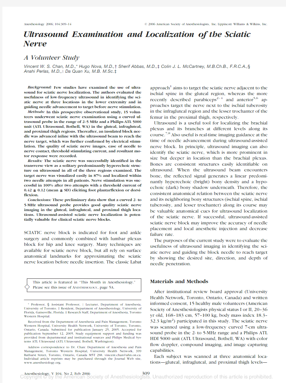
with the goal to localize the sciatic nerve at the level of the ischial spine,ischial tuberosity,and lesser trochan-ter,respectively.To scan the sciatic nerve in the ?rst two locations,the subject was positioned semiprone (Sim’s position)with the hip ?exed at approximately 90°.To scan in the proximal thigh,the probe was posi-tioned approximately 8cm distal to the inguinal crease,and the subject was supine with the hip and knee ?exed and the leg externally rotated at approximately 45°.The ultrasound probe was positioned perpendicular to the skin and oriented at each location to obtain the best possible transverse cross-sectional view of the sciatic nerve (i.e.,the ultrasound beam perpendicular to the nerve)as shown in ?gures 1A–C.
The following assessments were made at each anatom-ical location:the quality of ultrasound sciatic nerve im-ages (good image ?nerve recognized within 10s by two independent investigators,one performing ultrasound scanning and one independent observer;poor image ?one in which the nerve cannot be identi?ed by one or both investigators with con?dence and con?rmation re-quired nerve stimulation);nerve diameter (medial to lateral measurement);minimum skin-to-nerve distance (reported as mean ?SD);and identi?cation of neighbor-ing vascular,muscular,and bony structures.
After ultrasound scanning,a block needle was inserted for nerve localization and electrical stimulation in 15subjects.That is,each subject received two needle punc-tures (?rst,in the ischial spine location in all subjects,and second,in the ischial tuberosity location [8subjects]or the lesser trochanter location [7subjects]according to a computer-generated randomization table).Subse-quent electrical stimulation con?rmed target sciatic
nerve at both locations.After sterile skin preparation with povidone-iodine and local in?ltration with 1%lido-caine,an 8-cm,22-gauge insulated block needle (Pajunk,Geisingen,Germany)was inserted parallel and inline with the ultrasound probe covered with a sterile plastic cover and gel (?g.1D).The needle was advanced under real-time ultrasound imaging guidance until it made con-tact with the target nerve.When the needle was judged to be in satisfactory position,a nerve stimulator with a 100-?s pulse duration (Stimuplex;Braun Medical,Beth-lehem,PA)was turned on to elicit foot plantar?exion or dorsi?exion using a maximum of 1.5mA.After electrical stimulation,10–20ml dextrose 5%solution was injected incrementally in 2-to 3-ml aliquots under ultrasound observation to mimic a local anesthetic injection.The pattern of 5%dextrose solution spread,and the thresh-old stimulating current (mean ?SD)was noted.
Results
All subjects successfully completed the study.In the transverse view,the sciatic nerve appeared predomi-nantly hyperechoic on ultrasound as a ?at and thin ellip-tical structure at the gluteal region (ischial spine level,?g.2A),lip-to oval-shaped at the infragluteal region (ischial tuberosity level,?g.2B),and round or oval-shaped at the proximal thigh (lesser trochanter level,?g.2C).Good quality images of the sciatic nerve were ob-tained in 13of 15subjects (87%)at the gluteal region (ischial spine level)and 100%in the other two locations.When scanned obliquely in the buttock at the ischial spine level (?g.1A),the hyperechoic sciatic nerve was consistently located deep to the gluteal maximus
mus-
Fig. 1.Ultrasound probe positioned at three anatomical positions.It is posi-tioned oblique in the gluteal region of a subject lying semiprone (A )(GT ?greater trochanter;PSIS ?posterior su-perior iliac spine;SH ?sacral hiatus);transverse in the infragluteal region of a subject lying semiprone (B )(GT ?greater trochanter;IT ?ischial tuberos-ity);oblique and medially in the proxi-mal thigh of a subject lying supine,hip and knee ?exed and leg externally ro-tated (C )(IC ?inguinal crease);and in the proximal thigh with a needle passing inline with the ultrasound probe (D ).
310CHAN ET AL.
cle,lateral to the ischial spine,and above the hypere-choic ischial bone (?g.2A).The sciatic nerve was 1.67?0.17cm (mean ?SD)in width (medial to lateral mea-surement)and 3.48?0.91cm from the skin surface on ultrasound.Often,the pulsatile inferior gluteal artery pudendal artery,or both could be found medial to the sciatic nerve (?g.3A).
At the ischial tuberosity level,ultrasound scanning in the transverse plane (?g.1B)consistently showed the ischial tuberosity medially and greater trochanter later-ally,both hyperechoic in appearance (?g.2B).In be-tween was the hyperechoic oval-or lip-shaped sciatic nerve deep to the gluteal maximus muscle,most com-monly 1.57?0.17cm (mean ?SD)in width and 3.34?0.85from the skin surface.No nearby vessel was iden-ti?ed.
At the lesser trochanter level,when scanned approxi-mately 8cm distal to the inguinal crease and
transverse
Fig.2.Transverse sonograms of the sciatic nerve showing the hyperechoic nerve (arrowhead )at the ischial spine level (A )(A ?internal pudendal artery;GMM ?gluteus maximus muscle;IB ?ischial bone;IS ?ischial spine;V ?internal pudendal vein),the ischial tuberosity level (B )(GMM ?gluteus maximus muscle;GT ?greater trochanter;IT ?ischial tuberosity),and the lesser trochanter level (C )(AMM ?adductor magnus mus-cle;FA ?femoral artery;LT ?lesser
trochanter).
Fig.3.Transverse sonograms of the sciatic nerve with color doppler showing the nerve (arrowhead )in relation to the pu-dendal artery (red ,A)and vein (blue ,V)that are medial to the nerve at the ischial spine level (A )and in relation to the femoral artery (red ,FA)that is lateral to the nerve at the lesser trochan-ter level (B ).GMM ?gluteus maximus muscle;IB ?ischial bone;IS ?ischial spine;LT ?lesser trochanter.
311
ULTRASOUND-GUIDED SCIATIC NERVE LOCALIZATION
on the medial side of the thigh that was semi?exed and externally rotated (?g.1C),the hyperechoic sciatic nerve was found posterior and medial to the lesser tro-chanter (?g.2C).The nerve appeared most commonly oval or round,1.66?0.21cm in width and 6.21?0.68cm (mean ?SD)from the skin surface.The sciatic nerve was deep to the adductor magnus muscle,and the femoral neurovascular bundle was noted far lateral to the sciatic nerve in this projection (?g.3B).
Under real-time ultrasound guidance,all 15subjects underwent nerve localization and stimulation success-fully in two anatomical locations,ischial spine (n ?15)plus ischial tuberosity (n ?8)or lesser trochanter (n ?7).When the block needle was passed inline with the ultrasound beam (?g.1D),needle advancement was most commonly observed as needle and tissue (muscle)movement without clear view of the needle shaft due to the steep angle of needle penetration.We observed needle movement in real time until the needle made contact with the hyperechoic sciatic nerve as indicated by nerve movement.The needle-to-nerve distance was further minimized by adjusting the needle position based on nerve stimulation response.
When in contact with the sciatic nerve,electrical stim-ulation through the needle evoked muscle contraction in all cases as a con?rmatory signal.The minimum thresh-old currents required for nerve stimulation were 0.46?0.14,0.42?0.08,and 0.39?0.16mA at the level of the ischial spine,ischial tuberosity,and lesser trochanter,respectively,to elicit plantar?exion or dorsi?exion of the foot.Injection of 1–2ml of the 5%dextrose solution intensi?ed the motor response,and solution spreading around the nerve was observed after 10–20ml of injection (?g.4).There was no complication except mild buttock and posterior thigh paresthesia in one subject that lasted for 3days after the study.Paresthe-sia was not reported during needle advancement in any subject.
Discussion
Currently,high-resolution sonography is valuable in the diagnosis of peripheral nerve entrapment syn-dromes,9neuropathies,10and nerve tumors.11,12Previ-ous human and cadaver studies have examined anatomi-cal–sonographic correlation of the sciatic nerve,but they are limited to the mid thigh and regions be-low.10,13–17In this study,we have generated unique ultrasound images of the sciatic nerve in deeper loca-tions,i.e.,the gluteal,infragluteal,and proximal thigh regions.We have demonstrated that a low-frequency ultrasound probe in the range of 2–5MHz is valuable in localizing the sciatic nerve in these locations and can visually guide needle advancement to target with preci-sion.Ultrasound identi?cation of deep bony land-marks—ischial spine,ischial tuberosity,and lesser tro-chanter—provides a consistent guide to the sciatic nerve at these locations.Ultrasound-guided sciatic nerve block is potentially valuable for achieving clinical anesthesia with fewer attempts and higher success,but this needs con?rmation in future studies.
Labat’s approach to the sciatic nerve relies on inter-section of lines adjoining palpable surface bony land-marks (posterior superior iliac spine and the sacral hiatus medially and greater trochanter laterally).1The point of intersection indicates the approximate sciatic nerve lo-cation and the gluteal site of needle insertion.Anatomi-cally,the ischial spine is a more reliable bony landmark in proximity to the sciatic nerve,but it is not palpable on the skin surface.Chang et al.18described a technique of sciatic nerve block by palpating and identifying the is-chial spine directly upon rectal examination.Although this technique achieved block success in 76%of the cases,this method of sciatic nerve localization has not gained popularity for obvious reasons.
Our preliminary experience with ultrasound identi?-cation of the ischial spine is extremely encouraging.The imaging technique we use closely resembles that re-ported by Kovacs et al.,19who developed an ultrasono-graphic technique for pudendal nerve in?ltration for perineal pain.A 3.5-MHz curved-array probe was used to ?rst identify the ischial spine before localizing the pu-dendal nerve and internal pudendal artery located more medially.The internal pudendal artery (a branch of the internal iliac artery)was found in 98%of the cases and consistently within 1cm from the ischial spine.Our experience compares favorably with the ?ndings by Ko-vacs et al.Both the ischial spine and the inferior gluteal or internal pudendal artery (both branches of the inter-nal iliac artery)generate easily identi?able bony and vascular signals on ultrasound and are reliable cues me-dial to the sciatic nerve location (?gs.2A and
3A).
Fig.4.Transverse sonogram at the ischial spine level showing the sciatic nerve (arrowhead )after injection of 5%dextrose solution (D5W).GMM ?gluteal maximus muscle;IB ?ischial bone.
312CHAN ET AL.
Our?nding is also consistent with previous attempts by Hullander et al.,20who used Doppler ultrasound to detect superior gluteal arterial pulsation to aid sciatic nerve localization with the Labat approach.Both gluteal arteries are medial to the sciatic nerve with the superior gluteal artery passing between the L5and S1nerve roots and emerging from the upper border of the piriformis muscle while the inferior gluteal artery passes between S1and S2nerve roots and emerges below the piriformis muscle.
The infragluteal parabiceps technique2–4is a simple approach to the sciatic nerve where the nerve lies half-way between two easily identi?able bony landmarks,the greater trochanter laterally and the ischial tuberosity medially.A case of ultrasound-guided infragluteal sciatic nerve block was reported by Gray et al.21in a child undergoing Achilles tendon surgery.The block was suc-cessful,con?rmed with nerve stimulation,and local an-esthetic spread was observed surrounding the hypere-choic sciatic nerve deep to the gluteus maximus and
biceps femoris muscles.In the current study,we consis-tently observed enlargement of the presumably“sheath”compartment and extensive spreading of the injected5% dextrose solution completely or partially encircling the sciatic nerve within this compartment.We chose to pass the needle inline with the ultrasound beam as opposed to the perpendicular direction21so that needle advance-ment could be observed in real time.
The anterior approach as described by Beck5and Chelly and Delaunay6is to block the sciatic nerve at the lesser trochanter level of the femur.Direct needle pas-sage to the nerve is often blocked at this level when the leg is positioned supine and neutral.In a cadaver study, Vloka et al.22found that internal rotation of the leg by 45°greatly facilitated but external rotation consistently blocked needle passage at the lesser trochanter level. Alternatively,it is advantageous to block the sciatic nerve below the lesser trochanter because it is more accessible to needle passage when the leg is positioned supine and neutral.23,24
We took an entirely different approach in this study. When the thigh and knee are?exed and the leg is externally rotated(?g.1C),we?nd the sciatic nerve easily visible and accessible,posterior and deep to the femur at the lesser trochanter level and below.Further-more,the femoral neurovascular bundle is far lateral to the site of needle insertion;therefore any uninten-tional puncture is unlikely(?gs.2C and3B).In con-trast,ultrasound scanning directly over the femur with the leg in the conventional supine and neutral position showed the quadriceps muscles and the femur but not the sciatic nerve at the lesser trochanter level(?g.5), and the femoral neurovascular bundle is located me-dially.
In summary,ultrasound technology can provide high-quality images of the sciatic nerve and guide nerve lo-calization and needle placement in the gluteal,infraglu-teal,and thigh regions.Future studies are required to determine the clinical utility of ultrasound-assisted sci-atic nerve blocks in these locations.
References
1.Bridenbaugh PO,Wedel DJ:The lower extremity:Somatic blockade,Neural Blockade in Clinical Anesthesia and Management of Pain,3rd edition.Edited by Cousins MJ,Bridenbaugh PO.Philadelphia,Lippincott–Raven,1998,pp373–94
2.Sutherland ID:Continuous sciatic nerve infusion:Expanded case report describing a new approach.Reg Anesth Pain Med1998;23:496–501
3.Sukhani R,Candido KD,Doty R Jr,Yaghmour E,McCarthy RJ:Infragluteal-parabiceps sciatic nerve block:An evaluation of a novel approach using a single-injection technique.Anesth Analg2003;96:868–73
4.Di Benedetto P,Casati A,Bertini L,Fanelli G:Posterior subgluteal approach to block the sciatic nerve:Description of the technique and initial clinical experiences.Eur J Anaesthesiol2002;19:682–6
5.Beck GP:Anterior approach to sciatic nerve block.A NESTHESIOLOGY1963; 24:222–4
6.Chelly JE,Delaunay L:A new anterior approach to the sciatic nerve block.
A NESTHESIOLOGY1999;91:1655–60
7.Perlas A,Chan VW,Simons M:Brachial plexus examination and localization using ultrasound and electrical stimulation:A volunteer study.A NESTHESIOLOGY 2003;99:429–35
8.De Andres J,Sala-Blanch X:Ultrasound in the practice of brachial plexus anesthesia.Reg Anesth Pain Med2002;27:77–89
9.Delfaut EM,Demondion X,Bieganski A,Thiron MC,Mestdagh H,Cotten A: Imaging of foot and ankle nerve entrapment syndromes:From well-demonstrated to unfamiliar sites.Radiographics2003;23:613–23
10.Heinemeyer O,Reimers CD:Ultrasound of radial,ulnar,median,and sciatic nerves in healthy subjects and patients with hereditary motor and sensory neuropathies.Ultrasound Med Biol1999;25:481–5
11.Benyahya E,Etaouil N,Janani S,Bennis R,Tarfeh M,Louhalia S,Mkinsi O: Sciatica as the?rst manifestation of a leiomyosarcoma of the buttock.Rev Rhum Engl Ed1997;64:135–7
12.Dubuisson A,Fissette J,Vivario M,Reznik M,Stevenaert A:A benign tumor of the sciatic nerve:Case report and review of the literature.Acta Neurol Belg 1991;91:5–11
13.Fornage BD:Peripheral nerves of the extremities:Imaging with US.Radi-ology1988;167:179–82
14.Peer S,Kovacs P,Harpf C,Bodner G:High-resolution sonography of lower extremity peripheral nerves:Anatomic correlation and spectrum of disease.J Ultrasound Med2002;21:315–22
15.Graif M,Seton A,Nerubai J,Horoszowski H,Itzchak Y:Sciatic nerve: Sonographic evaluation and anatomic-pathologic considerations.Radiology1991; 181:405–8
16.Sinha A,Chan VW:Ultrasound imaging for popliteal sciatic nerve block. Reg Anesth Pain Med2004;
29:130–4
Fig.5.Transverse sonogram at the lesser trochanter(LT)level with the leg in a supine neutral position.The sciatic nerve being posterior to the femur is not visualized in this projection.The femoral artery is located medially(red,FA)when the leg is positioned neutral.
313
ULTRASOUND-GUIDED SCIATIC NERVE LOCALIZATION
17.Sites BD,Gallagher J,Sparks M:Ultrasound-guided popliteal block dem-onstrates an atypical motor response to nerve stimulation in2patients with diabetes mellitus.Reg Anesth Pain Med2003;28:479–82
18.Chang PC,Lang SA,Yip RW:Reevaluation of the sciatic nerve block.Reg Anesth1993;18:18–23
19.Kovacs P,Gruber H,Piegger J,Bodner G:New,simple,ultrasound-guided in?ltration of the pudendal nerve:ultrasonographic technique.Dis Colon Rectum 2001;44:1381–5
20.Hullander M,Spillane W,Leivers D,Balsara Z:The use of Doppler ultra-sound to assist with sciatic nerve blocks.Reg Anesth1991;16:282–4
21.Gray AT,Collins AB,Schafhalter-Zoppoth I,:Sciatic nerve block in a child:
A sonographic approach.Anesth Analg2003;97:1300–2
22.Vloka JD,Hadzic A,April E,Thys DM:Anterior approach to the sciatic nerve block:The effects of leg rotation.Anesth Analg2001;92:460–2
23.Van Elstraete AC,Poey C,Lebrun T,Pastureau F:New landmarks for the anterior approach to the sciatic nerve block:Imaging and clinical study.Anesth Analg2002;95:214–8
24.Ericksen ML,Swenson JD,Pace NL:The anatomic relationship of the sciatic nerve to the lesser trochanter:Implications for anterior sciatic nerve block.Anesth Analg2002;95:1071–4
314CHAN ET AL.
坐骨神经痛最好的治疗方法
坐骨神经痛最好的治疗方 法 This model paper was revised by the Standardization Office on December 10, 2020
坐骨神经痛最好的治疗方法 坐骨神经痛是指坐骨神经病变,沿坐骨神经通路即腰、臀部、大腿后、小腿后外侧和足外侧发生的疼痛症状群,坐骨神经是支配下肢的主要神经干,坐骨神经痛是指坐骨神经通路及其分布区域内(臀部、大腿后侧、小腿后外侧和脚的外侧面)的疼痛。本病男性青壮年多见,单侧为多,疼痛程度及时间常与病因及起病缓急有关。 坐骨神经痛的临床表现: 1、根性坐骨神经痛:起病随病因不同而异。最常见的腰椎间盘突出,常在用力、弯腰或剧烈活动等诱因下,急性或亚急性起病。少数为慢性起病。疼痛常自腰部向一侧臀部、大腿后,腘窝、小腿外侧及足部放射,呈烧灼样或刀割样疼痛,咳嗽及用力时疼痛可加剧,夜间更甚。病员为避免神经牵拉、受压,常取特殊的减痛姿势,如睡时卧向健侧,髋、膝关屈曲,站立时着力于健侧,日久造成脊柱侧弯,多弯向健侧,坐位进臀部向健侧倾斜,以减轻神经根的受压。 2、干性坐骨神经痛:起病缓急也随病因不同而异。如受寒或外伤诱发者多急性起病。疼痛常从臀部向股后、小腿后外侧及足外侧放射。行走、活动及牵引坐骨神经时疼痛加重。压痛点在臀点以下,Lasegue征阳性而Kernig征多阴性,脊椎侧弯多弯向患侧以减轻对坐骨神经干的牵拉。
坐骨神经痛的危害: 目前,大部分人都将坐骨神经痛作为一种病来看待,专家王怀庆解释说其实这是不科学的。坐骨神经痛并不是一种病,而是一种由其他疾病引起的症状,危害是非常大的。原发性坐骨神经痛多于受寒和感染有关,会和肌炎、肌纤维组织炎等一起产生,以单侧为多见的,严重的可能致残。而继发性坐骨神经痛大都是由于临近结构的病变所引起的,患者也会疼痛难忍,久而久之会造成脊柱侧弯,影响到正常的站姿和坐姿。 坐骨神经痛的治疗方法: 坐骨神经痛西药久治不愈,是腰椎病变压迫导致,职业病的表现。中医遵循通则不痛的原理,采用外敷 治、作用持久,可以达到疏通经络,消肿止痛的作用,能从根本调治坐骨神经痛。 坐骨神经痛的运动疗法: 坐骨神经痛多发于单侧 ,夜间加重 ,咳嗽、大便时加重。坐骨神经痛分为原发性和继发性两种。原发性的主要是由于坐骨神经炎症病变引起 ;继发性的则多由腰椎间盘突出症、腰椎增生、腰和臀部的软组织损伤以及盆腔、椎管内病变引起。患者除避免着凉外 ,适当加强腰腿部功能锻炼 ,会获得良好效果。
腰椎间盘突出特效方 (绝对秘方)
腰椎间盘突出特效方(绝对秘方)绝对秘方(腰突、坐骨神经痛) 此方为旧社会本地一地主家中所藏。相传古时地主家中有人患腰腿痛久治不效,后重金请名医岳先生开出此方,配成药酒一剂而愈。地主把此方写在墙上,流传至今。此方在当地流传甚广,我十年前在地主后人处偶然得到此方,看方子组成简单并无甚出奇之处,在遇到不愿长期吃苦药的病人时开出此方,经治不少病人有效者竟占7成左右。今贴方于此有兴趣的朋友不妨一试!(腰椎管狭窄、退行性脊柱炎、腰椎间盘突出症、梨状肌综合症及不明原因的坐骨神经痛皆可用之) 组成: 当归12g 川芎12g 木瓜12g 牛膝12g 红花6g 大力草12g 炙乳香 12g 全虫70个肉桂15g 杜仲9g 二花15g 乌梅12g 陈皮12g 甘草15g 用水3斤煎熬后滤出药汁2斤左右加入白酒2斤,红糖2两、白糖2两。装入干净容器中保存。每次服1两,每日3次。 民间流传的腰椎间盘突出症方一副就好? 今天临县有个病人拿个方子来找我,让我看看能否服用,说她弟弟的腰突症几年前就是吃这个方子好的,并说方子很神!(家乡周围吃愈了好多人)。方子我贴上来让大家瞧瞧! 生川乌25g 当归15g 木瓜15g 砂仁15g 明雄10g 栀子15g 川牛膝 15g 广木香15g 粉碎泛为水丸或装胶囊,大号胶囊每日三次、每次一粒。 腰突良方——药物渗透 腰突良方 去年有一教师腰疼的不敢站立,X光片示L4,5椎间盘突出,我单用五川灵仙汤九天后,X光片示正常片,后来我用此方,治疗颈肩腰腿疼也取得满意效果.具体操作是:药物组成:川芎20克,川乌20克,川牛膝30克,川断30克,川椒20克,威灵仙30克,木瓜20克,透骨草30克,鸡血藤30克,玄胡20克,乳香20克,没药20克,芒硝(另包)50克,食醋250毫升。 临床上我把除芒硝,食醋外的药装到布袋中,把布袋放到微波
腰椎间盘突出特效方绝对秘方
腰椎间盘突出特效方绝 对秘方 TTA standardization office
腰椎间盘突出特效方绝 对秘方 Pleasure Group Office【T985AB-B866SYT-B182C-BS682T-STT18】
腰椎间盘突出特效方(绝对秘方)绝对秘方(腰突、坐骨神经痛) 此方为旧社会本地一地主家中所藏。相传古时地主家中有人患腰腿痛久治不效,后重金请名医岳先生开出此方,配成药酒一剂而愈。地主把此方写在墙上,流传至今。此方在当地流传甚广,我十年前在地主后人处偶然得到此方,看方子组成简单并无甚出奇之处,在遇到不愿长期吃苦药的病人时开出此方,经治不少病人有效者竟占7成左右。今贴方于此有兴趣的朋友不妨一试!(腰椎管狭窄、退行性脊柱炎、腰椎间盘突出症、梨状肌综合症及不明原因的坐骨神经痛皆可用之)组成:当归12g川芎12g木瓜12g牛膝12g红花6g大力草12g炙乳香12g全虫70个肉桂15g 杜仲9g 二花15g乌梅12g陈皮12g甘草15g 用水3斤煎熬后滤出药汁2斤左右加入白酒2斤,红糖2两、白糖2两。装入干净容器中保存。每次服1两,每日3次。 民间流传的腰椎间盘突出症方一副就好 今天临县有个病人拿个方子来找我,让我看看能否服用,说她弟弟的腰突症几年前就是吃这个方子好的,并说方子很神!(家乡周围吃愈了好多人)。方子我贴上来让大家瞧瞧! 生川乌25g 当归15g木瓜15g砂仁15g明雄10g栀子15g川牛膝15g广木香15g 粉碎泛为水丸或装胶囊,大号胶囊每日三次、每次一粒。 腰突良方——药物渗透 腰突良方 去年有一教师腰疼的不敢站立,X光片示L4,5椎间盘突出,我单用五川灵仙汤九天后,X光片示正常片,后来我用此方,治疗颈肩腰腿疼也取得满意效果.具体操作是:药物组成:川芎20克,川乌20克,川牛膝30克,川断30克,川椒20克,威灵仙30克,木瓜20克,透骨草30克,鸡血藤30克,玄胡20克,乳香20克,没药20克,芒硝(另包)50克,食醋250毫升。
坐骨神经痛的症状图片
坐骨神经痛的症状图片 坐骨神经从腰椎脊髓发出,沿着臀部和腿部一路到达脚部,是人体最大最长的一根神经。大概分为三种情况,有一种是因为外部的创伤,或者是因为受凉引起的疼痛,会偶然间的痛一下,疾病治疗也很容易复发,大多数情况下都是一侧有疼痛的感觉,很少有双侧都疼痛的情况。再有就是全身性的疼痛,表现症状是背部腰部,逐渐感觉僵硬麻木,疼痛的感觉逐渐加重,慢慢引发到腰部臀部等地方,最后大腿后侧小腿外侧,包括主妇都会有痛的感觉,阵发性的疼痛,在半夜疼痛会更厉害一点。 坐骨神经痛的治疗方法最好选用保守的方法恢复:平时注意休息,注意防护,不要剧烈运动,避免劳累,多吃蔬菜水果,外用掏、宝的(济愈堂坐骨顺古安玉贴),修复坐骨神经病变,相对于传统的方式,坐骨贴有以下优势: 1. 延长给药时间维持恒定的有效药物浓度,提高疗效,达到良好的治疗效果。 2.药物不经消化道,避免胃酸对药效的破坏作用及对胃肠道的刺激。 3. 不损伤肝肾,药物是通过皮肤穴位吸收,不走肝脏,用的又是中药,避免了对肝肾的潜在危险。 4. 操作简单,治疗用时短,不耽搁病人工作。
5.无痛苦、无伤害,远离抗生素,减少打针、输液带来的痛苦及危害。 坐骨神经痛有什么症状: 1、疼痛主要限于坐骨神经分布区,大腿后部、小腿后外侧和足部,疼痛剧烈的病人可呈特有的姿势;腰部屈曲、屈膝、脚尖着地。如病变位于神经根时,椎管内压力增加时疼痛加重。 2、肌力减退的程度可因病因、病变部位、损害的程度不同差异很大,可有坐骨神经支配肌肉全部或部分力弱或瘫痪。 3、可有或无坐骨切迹处坐骨神经干的压痛。 4、有坐骨神经牵拉征,及其等位征阳性,此征的存在常与疼痛的严重程度相平行。局麻坐骨神经根或神经干此征可消失。 5、跟腱反射减退或消失,膝反射可因刺激而增高。 6、可有坐骨神经支配区域的各种感觉的减退或消失,包括外踝的振动觉减退,亦可有极轻的感觉障碍。
绝对秘方(腰突、坐骨神经痛)
[转载]绝对秘方(腰突、坐骨神经痛) (2012-10-29 01:02:21) 转载▼ 标签: 转载 原文地址:绝对秘方(腰突、坐骨神经痛)作者:闲在居士 作者:混元整体 此方为旧社会本地一地主家中所藏。相传古时地主家中有人患腰腿痛久治不效,后重金请名医岳先生开出此方,配成药酒一剂而愈。地主把此方写在墙上,流传至今。此方在当地流传甚广,我十年前在地主后人处偶然得到此方,看方子组成简单并无甚出奇之处,在遇到不愿长期吃苦药的病人时开出此方,经治不少病人有效者竟占7成左右。今贴方于此有兴趣的朋友不妨一试!(腰椎管狭窄、退行性脊柱炎、腰椎间盘突出症、梨状肌综合症及不明原因的坐骨神经痛皆可用之)
组成: 当归12g 川芎12g 木瓜12g 牛膝12g 红花6g 大力草12g 炙乳香 12g 全虫70个肉桂15g 杜仲9g 二花15g 乌梅12g 陈皮12g 甘草15g 用水3斤煎熬后滤出药汁2斤左右加入白酒2斤,红糖2两、白糖2两。装入干净容器中保存。每次服1两,每日3次。 民间流传的腰椎间盘突出症方一副就好? 今天临县有个病人拿个方子来找我,让我看看能否服用,说她弟弟的腰突症几年前就是吃这个方子好的,并说方子很神!(家乡周围吃愈了好多人)。方子我贴上来让大家瞧瞧! 生川乌25g 当归15g 木瓜15g 砂仁15g 明雄10g 栀子15g 川牛膝15g 广木香15g 粉碎泛为水丸或装胶囊,大号胶囊每日三次、每次一粒。 腰突良方——药物渗透
腰突良方 去年有一教师腰疼的不敢站立,X光片示L4,5椎间盘突出,我单用五川灵仙汤九天后,X光片示正常片,后来我用此方,治疗颈肩腰腿疼也取得满意效果.具体操作是:药物组成:川芎20克,川乌20克,川牛膝30克,川断30克,川椒20克,威灵仙30克,木瓜20克,透骨草30克,鸡血藤30克,玄胡20克,乳香20克没药20克,芒硝(另包)50克,食醋250毫升。 临床上我把除芒硝,食醋外的药装到布袋中,把布袋放到微波盒中,把芒硝放入适量温水中搅拌,尽量溶解,溶液倒如入微波盒中,再倒入微波盒适量温水,瞒过药袋,然后放到微波炉中,高火五分钟,取出微波盒把食醋倒入其中,再高火五分钟,取出,待袋子不是很烫后放到患处,然后在袋上盖上一塑料这样保温.一次半小时,一天两次.效果确实很好,大家试试吧!临床上我不单用于腰突,其中颈椎病,肩周炎,肋间神经疼
坐骨神经痛的锻炼图片
坐骨神经痛的锻炼图片 坐骨神经痛是指坐骨神经病变,坐骨神经痛症状主要是有沿坐骨神经通路即腰、臀部、大腿后、小腿后外侧和足外侧发生的疼痛症,坐骨神经痛的治疗方法主要是药物治疗。可应用掏宝的【济愈堂坐骨贴】注意避免过度劳累及剧烈运动,注意休息、可以平躺平板床。 坐骨神经痛有什么症状 1、疼痛主要限于坐骨神经分布区,大腿后部、小腿后外侧和足部,疼痛剧烈的病人可呈特有的姿势;腰部屈曲、屈膝、脚尖着地。如病变位于神经根时,椎管内压力增加时疼痛加重。 2、肌力减退的程度可因病因、病变部位、损害的程度不同差异很大,可有坐骨神经支配肌肉全部或部分力弱或瘫痪。 3、可有或无坐骨切迹处坐骨神经干的压痛。 4、有坐骨神经牵拉征,及其等位征阳性,此征的存在常与疼痛的严重程度相平行。局麻坐骨神经根或神经干此征可消失。 5、跟腱反射减退或消失,膝反射可因刺激而增高。 6、可有坐骨神经支配区域的各种感觉的减退或消失,包括外踝的振动觉减退,亦可有极轻的感觉障碍。
坐骨神经痛的锻炼图片 锻炼方法: 1、拱身如桥:躺在床上,把骨盆挺起来,像打挺似的两脚着床,上背和腰不要着床,就像一座独木桥似的,这样挺起来以后坚持十分钟,然后反复做5—10次,能够很好的锻炼骨盆中的骶椎。 2、小燕飞:趴着像小燕飞,挺起来,飞起来,双腿尽量向后翘,对骨盆有很好的治疗效果。 3、仰卧起坐:大家很熟悉这个动作,因为在做仰卧起坐时候,骶骨的骶尖部是顶在床上的,身体一下一下地坐起来,这个姿势有利于骶骨,使骶尖上翘向正确方向恢复。当然有的朋友说,这个仰卧起坐太费劲我做不了两个,甚至做完以后腰有点不舒服,这个问题很好解决,你要平躺着做仰
治疗骨病秘方
腰腿痛颈椎病健康 此方通治:风湿、类风湿、坐骨神经痛、肩周炎、颈椎病、骨质增生、四肢麻木、肌肉萎缩、股骨头坏死、椎间盘突出、腰腿痛等一切骨科疾病。 药方贡献人:洪真大居士 洪真居士;是佛门高德之士。出自中医世家,现任某医院院长一职。曾得多位佛、道两教高人真传,穷其一生研究......。好多,不多说。今有两门中医绝技(根治一切胃肠疾病\根治一切骨科疾病)临床应用效验如神,故此贡献于世。广利有情,救济世人,乃洪真居士之愿!也是我佛之愿!天下病苦众生之愿! 配方:麻黄10克、艾叶10克、乌梢蛇15克、蕲蛇15克、乳香10克、白芷10克、威灵仙50克、千年健10克、钻地风10克、伸筋草30克、鸡血藤30克、羌活15克、独活15克、牛夕15克、当归12克、防己12克、桂枝15克、木瓜12克、葛根15克、穿山甲15克、蚂蚁50克、黄芪20克、白术15克、一口钟80克金樱子15克、甘草15克。药店加工成粉。 服法:每天服3次,按各人病情轻重每次5到10克(一小汤勺或两小汤勺)三餐前半小时服用。一到三
副药可以根治 治高血压: (1)香蕉皮30克,晒干水煎喝,每日3次,一个月见效。(2)用中药罗布麻,开水冲喝,每日15克,半月见效。 (3)银杏叶每日15克,用开水冲喝下,半月见效。 治低血压: 甘草20克,桂枝,肉桂各40克,将以上药物混合后当茶冲泡服用一周。 治血脂稠: (1)枸杞子10克,何首乌,草决明,山楂各15克,丹参20克,水煎服,每日两次,四个疗程治愈,(七天为一个疗程)。 (2)山楂. 银杏叶. 绞股蓝各15克,泡茶喝。连服四疗程(半月为一疗程) 脸上黑星(雀斑): 元荽(又名香菜),煎汤,一天洗三次,一个疗程治愈。 治脚汗. 脚臭: 白萝卜煮水,每晚熏洗双脚30分钟,连洗半月治愈。 治脚气: 韭菜一斤,煮水十分钟泡脚,每日一次,每次20分钟,三天除根。
坐骨神经痛的健身锻炼
坐骨神经痛的健身锻炼 在家可适当做伸延运动来减少疼痛和预防坐骨神经痛,但是,因为人和人的不同,每个人的运动也有所不同。对我们大多数人来说,行走和游泳可强化后背肌肉。你坐着、站着和躺着的方式可能有重要影响。 如果长时间站立,你的头应该向前,你的背部应该挺直。均匀分配两脚重力,保持腿部直立。坐着时你的腰背部应该有支撑,背部保持伸直状态。臀部略高于膝部,让脊椎下部自然弯曲,给予神经活动的充足空间,脚应该平放于地面——如必要使用一个脚凳。如果感觉舒服的话,使用一个小垫子或者成卷的毛巾支撑腰背部。 过去,那些背痛的人被要求睡硬床垫,但是有研究显示,选择硬度适中的床垫最好。如果你的床垫太软,那就在床基上面和床垫下面放一个硬板。使用枕头支撑你的头部,但是,要确保你的颈部不会大角度上扬。 当然也与运气有关,你可能在错误的时间做错误的运动,最终导致背痛和坐骨神经痛。我们必须承认,只要运动我们的背部就容易疼痛,因为老化有些运动我们需要限制。最好的办法是保持自己的身体柔软和强壮并保持手指交叉。 抬腿,平躺于地板上,双臂置于两侧。轮流抬起每个脚后跟,使脚后跟抬离地面10到20厘米,与此同时,腿
保持伸直状态。坚持,直到不舒服为止。重复5次。 预防坐骨神经痛:沿墙滑动,靠墙站立,双脚分开与肩同宽,然后下滑,蹲伏,膝部弯曲约90度。从1数到5,然后上滑。重复5次。 预防坐骨神经痛:抬高臀部平躺在地板上,屈膝,使得脚平放在地面上。然后抬起臀部,胃肌绷紧,与此同时挺直背部。重复5次。 适当加强腰腿部功能锻炼,会获得良好效果。 左右摆腿。站立位,双手扶墙,轮流向左右方向摆腿,摆动时足部不触地面。 交替直腿上抬运动。仰卧位,轮流将在、右腿伸直后抬起,经常锻炼可逐渐提高抬举角度。 踏自行车运动。仰卧位,两下肢像骑车般轮番踩踏,踩踏幅度可逐渐增加。 正坐举腿。坐位,两腿紧靠或夹上一本厚书,直膝,脚跟着地,手握凳边,抬腿过脐,随即放下。开始时患腿未必抬得很高,坚持锻炼后患腿的抬高程度会逐渐增加。 平坐推腿。坐位,足跟着地,足尖跷起,两手平放大腿上,随即向前弯腰,两手同时推向足部。初练时两手很难推到足部,坚持一段时间会收到良好的效果。 蹲跳。双手扶凳,左腿屈膝下蹲,右腿尽量向右侧伸直,如此左右交替进行。
狗骨头泡酒治骨刺,民间治疗骨病小偏方
狗骨头泡酒治骨刺,民间治疗骨病小偏方 大家都应该知道狗是热性动物,其骨坚韧,俗话说炸不烂的狗头,以骨治骨,用狗骨治料类风湿病,实际上就是用狗骨的残存能量来补足人体亏虚的能量,这也是能量转移。 1.腿抽筋:桑椹子100克,煎水服,一日二次,5天即效。 2.四肢麻木:老丝瓜筋50克,煎水服,一日二次,连服7天,有特效。 3.骨刺(骨质增生):狗骨头300克,砸碎炒黄,白酒1500毫升,浸泡3天后,用狗骨酒擦患处,每次服狗骨酒30毫升,一日三次,连服用半月。 4.颈椎痛:羊骨头(生的、煮过的均可)300克,砸碎炒黄,白酒1500毫升,浸泡3天后,用羊骨酒擦颈部,一日三次,连用半月。 5.坐骨神经痛:食用盐500克(可反复使用),炒热后加艾叶30克,用布包好敷患处至盐凉,一日一次,连用10天。
6.落枕(睡觉时由于枕头或姿势不适,而引起的颈痛):韭菜汁加热擦颈部,日擦7次,3天可好。 7.关节炎、肩周炎(包括风湿性、类风湿性关节炎):食用盐500克,放锅内炒热,再加葱须、生姜各3钱,一起用布包好,趁热敷患处至盐凉。一日一次,连用7天,有追风祛湿功效。 8.劳伤腰痛:艾叶30克,炒黄的蟹壳30克,浸白酒500毫升,3日后用药酒涂腰部,一日三次,连用10天,多可治多年腰痛。 9.坐骨神经痛神效方:川乌10克、麻黄10克、羌活25克、黄芪25克、桂枝20克、甘草15克,水煎服,日一剂,分服三次。 10.滑膜炎方:栀子250克,研末,用白酒和成泥状,敷在膝盖处,然后缠纱布,怕渗漏可以缠一层保鲜膜。每晚睡觉时敷好,白天取下,连敷7天。 11.腰椎间盘突出方:蔓荆子、刘寄奴、红花、防风、木香、透骨草、甘草、独活、白芷、没药、牛蒡子、川断、姜活、生盐各60克,研末,加上生盐掺匀放在碗里蒸热,趁热用布包住烫患处,至凉时贴在患处,每天早晚一次,一剂分4贴。药干了时取下保存,反复蒸用,4天一剂药,用三剂多能痊愈。
坐骨神经痛最好的治疗方法
坐骨神经痛最好的治疗 方法 Document number:PBGCG-0857-BTDO-0089-PTT1998
坐骨神经痛最好的治疗方法 坐骨神经痛是指坐骨神经病变,沿坐骨神经通路即腰、臀部、大腿后、小腿后外侧和足外侧发生的疼痛症状群,坐骨神经是支配下肢的主要神经干,坐骨神经痛是指坐骨神经通路及其分布区域内(臀部、大腿后侧、小腿后外侧和脚的外侧面)的疼痛。本病男性青壮年多见,单侧为多,疼痛程度及时间常与病因及起病缓急有关。 坐骨神经痛的临床表现: 1、根性坐骨神经痛:起病随病因不同而异。最常见的腰椎间盘突出,常在用力、弯腰或剧烈活动等诱因下,急性或亚急性起病。少数为慢性起病。疼痛常自腰部向一侧臀部、大腿后,腘窝、小腿外侧及足部放射,呈烧灼样或刀割样疼痛,咳嗽及用力时疼痛可加剧,夜间更甚。病员为避免神经牵拉、受压,常取特殊的减痛姿势,如睡时卧向健侧,髋、膝关屈曲,站立时着力于健侧,日久造成脊柱侧弯,多弯向健侧,坐位进臀部向健侧倾斜,以减轻神经根的受压。 2、干性坐骨神经痛:起病缓急也随病因不同而异。如受寒或外伤诱发者多急性起病。疼痛常从臀部向股后、小腿后外侧及足外侧放射。行走、活动及牵引坐骨神经时疼痛加重。压痛点在臀点以下,Lasegue征阳性而Kernig征多阴性,脊椎侧弯多弯向患侧以减轻对坐骨神经干的牵拉。
坐骨神经痛的危害: 目前,大部分人都将坐骨神经痛作为一种病来看待,专家王怀庆解释说其实这是不科学的。坐骨神经痛并不是一种病,而是一种由其他疾病引起的症状,危害是非常大的。原发性坐骨神经痛多于受寒和感染有关,会和肌炎、肌纤维组织炎等一起产生,以单侧为多见的,严重的可能致残。而继发性坐骨神经痛大都是由于临近结构的病变所引起的,患者也会疼痛难忍,久而久之会造成脊柱侧弯,影响到正常的站姿和坐姿。 坐骨神经痛的治疗方法: 坐骨神经痛西药久治不愈,是腰椎病变压迫导致,职业病的表现。中医遵循通则不痛的原理,采用外敷 治、作用持久,可以达到疏通经络,消肿止痛的作用,能从根本调治坐骨神经痛。 坐骨神经痛的运动疗法: 坐骨神经痛多发于单侧 ,夜间加重 ,咳嗽、大便时加重。坐骨神经痛分为原发性和继发性两种。原发性的主要是由于坐骨神经炎症病变引起 ;继发性的则多由腰椎间盘突出症、腰椎增生、腰和臀部的软组织损伤以及盆腔、椎管内病变引起。患者除避免着凉外 ,适当加强腰腿部功能锻炼 ,会获得良好效果。
腰椎间盘突出腰突、坐骨神经痛秘方偏方
腰椎间盘突出(腰突)、坐骨神经痛秘方偏方 (故事)治腰突、腰痛的一种简捷实用根本办法 方法:用陈林峰先生所创的《反正法》之第三法,即“足根蹬108次,足尖蹬49次。蹬时意念集中于两足、颈椎至腰椎。站、坐卧均可”,反复再三用之,可愈也。 唯用此法时,不可蛮干;不可畏痛,而要用力屈伸,力达而止;勤而习之,必可成功。 待膀胱经通畅之时,诸痛必无矣。正所谓“通则不痛,痛则不通;不通则痛,不痛则通。” ====================================== ====================================== ========= 老张突然腰痛难忍,经医学影像确诊为腰椎间盘突出,腰4-5右侧,症见腰痛及臀部放射痛,经多方求治不曾痊愈。后经一位老太太指点,用一个单方治好了。
配方:白面 白酒 各适量。用法:用白酒搅拌白面成糊状,涂糊在患部,昼夜不停,面干了就再涂糊,到三四天时,痒的难受,为防止手抓瘙痒感染,使用火罐拔局部,拔完再糊,糊完再拔,连续治疗半个月,疼痛诸症消失,活动自如,不会复发。腰间盘突出,涂酒拌面糊,治疗半个月,痛消如当初。【杨氏诊所原创】 [转载]神了:黄芪外洗根除腰椎间盘突出症 笔者在实践中发现,应用黄芪洗液可根除腰椎间盘突出症。且花费低廉,效果好,无副作用。具体做法如下: 1.用量:黄芪250克,计量加大后有刺激性。如果患者可以承受的话也可以逐步加量。 2.制备:使用铝锅和煤气灶,将黄芪制成足够洗浴的汤剂(该
药易沸,煎时小心)。洗完后,如有条件可将药液灌入桶内,放到冰箱里,留待下一次再用,一般可反复使用3—10次。 3.用法:趁热洗浴,总时间约一小时左右,选用一些保温措施保持室内温度,并轮流加热一部分洗液,总体以舒适为宜。必须使有病部位浸到药液,腰痛浸腰,腿痛浸腿。如果有肩周炎或颈椎病也要用水洗到,该方只对洗过的部位有效。 4.疗效:使用第一次后,即可松弛紧张的肌肉,提高患病部位的体温,祛除疼痛;使用5天后,如发现存有不能治疗的症状(如腰椎错位),找医生复查或纠正一下即可;使用15天后即可认为根除病症,病人也可根据实际情况多洗几天。全程不要求病人停止工作,病人可适量提重物;暂时未发现戒口,但病人营养好一些可能会有益。由于病患的全息相关性,存在脏腑辨证的胃病、妇科病等可一并治愈。 笔者认为,黄芪有补气利水的功效,热浴可以活血,该方通过改善局部血运来治病,印证了中医“通则不痛”的观点。------------------------------------------ 以下所列方药,均来自乌梅 的博客。 1
坐骨神经疼的位置图片
坐骨神经疼的位置图片 坐骨神经痛是以坐骨神经径路及分布区域疼痛为主的综合征。坐骨神经痛的绝大多数病例是继发于坐骨神经局部及周围结构的病变对坐骨神经的刺激压迫与损害,称为继发坐骨神经痛;少数系原发性,即坐骨神经炎。 方法/步骤 1 坐骨神经痛症状: 病人腰部不能直立,他要屈膝、屈髋,走路的时候脚尖沾地,脚部不敢完全踩实 2 坐骨神经痛原因: 腰源性的:腰椎间盘突出,腰椎的骨性关节炎,腰椎管狭窄,骶髂关节的损伤,骶髂关节炎非腰源性的:坐骨神经走形部位周边的组织,压迫坐骨神经3 坐骨神经痛病人腰部反应: 病因是腰源性的,会腰部疼痛病因不是腰源性的,腰部疼痛会轻一些 4 坐骨神经痛治疗方法:
对因治疗,如果病因是腰源性的,比如是腰椎间盘突出,就要按腰椎间盘突出的治疗方法如果病因是周围组织卡压,就要搞清楚周围组织的原因,把肌肉放松或者把炎症解除,坐骨神经疼就会明显的缓解 5 中药: 使用济愈堂坐骨顺古安玉贴,修复坐骨神经病变。可以达到摆脱坐骨神经疼的目的。 6 卧床休息注意事项: 卧床休息比较有效缓解疼痛的比较典型的一个姿势就是膝肘卧位:趴在床上屈膝,然后曲肘,弓着趴在床上这样能够把坐骨神经的神经孔放到最大的一个程度,这样时压力最小,所以说这种姿势能够非常有效的缓解坐骨神经疼7 坐骨神经疼的自我康复方法: 坐骨神经疼的急性期缓解了以后,可以适当的做点轻度的体育锻炼做做腰椎的保健操,可以恢复腰部和腿部的肌肉力量,达到一个比较好的预防作用 坐骨神经痛的日常护理: 1 防止吹太久的风,夏天天气炎热,大量的汗液外流,风扇,空调的长时间吹袭,很容易就导致坐骨神经痛。所以,即使是在炎热的夏天,也不可贪凉哦。 2 防寒,冬天要时刻注意保暖,晚上睡觉时要防止肩关节外露。 3 避免外伤日常生活要注意安全,避免受伤。在运动受伤之后要及时医治。 4 多平躺休息,平躺在床上可以减轻神经和病变组织的张力和水肿,以此缓解坐骨神经痛。当病情得到缓解,在适当的运动运动。 5 针对病因进行治疗,疼痛比较严重的患者,需要对症下药。
坐骨神经痛的症状和治疗方法
坐骨神经痛的症状和治疗方法 坐骨神经从腰椎脊髓发出,沿着臀部和腿部一路到达脚部,是人体最大最长的一根神经。大概分为三种情况,有一种是因为外部的创伤,或者是因为受凉引起的疼痛,会偶然间的痛一下,疾病治疗也很容易复发,大多数情况下都是一侧有疼痛的感觉,很少有双侧都疼痛的情况。再有就是全身性的疼痛,表现症状是背部腰部,逐渐感觉僵硬麻木,疼痛的感觉逐渐加重,慢慢引发到腰部臀部等地方,最后大腿后侧小腿外侧,包括主妇都会有痛的感觉,阵发性的疼痛,在半夜疼痛会更厉害一点。 坐骨神经痛的治疗方法最好选用保守的方法恢复:平时注意休息,注意防护,不要剧烈运动,避免劳累,多吃蔬菜水果,外用掏、宝的(坐骨顺古安玉贴),修复坐骨神经病变,相对于传统的方式,坐骨贴有以下优势: 1. 延长给药时间维持恒定的有效药物浓度,提高疗效,达到良好的治疗效果。 2.药物不经消化道,避免胃酸对药效的破坏作用及对胃肠道的刺激。 3. 不损伤肝肾,药物是通过皮肤穴位吸收,不走肝脏,用的又是中药,避免了对肝肾的潜在危险。 4. 操作简单,治疗用时短,不耽搁病人工作。
5.无痛苦、无伤害,远离抗生素,减少打针、输液带来的痛苦及危害。 坐骨神经痛有什么症状: 1、疼痛主要限于坐骨神经分布区,大腿后部、小腿后外侧和足部,疼痛剧烈的病人可呈特有的姿势;腰部屈曲、屈膝、脚尖着地。如病变位于神经根时,椎管内压力增加时疼痛加重。 2、肌力减退的程度可因病因、病变部位、损害的程度不同差异很大,可有坐骨神经支配肌肉全部或部分力弱或瘫痪。 3、可有或无坐骨切迹处坐骨神经干的压痛。 4、有坐骨神经牵拉征,及其等位征阳性,此征的存在常与疼痛的严重程度相平行。局麻坐骨神经根或神经干此征可消失。 5、跟腱反射减退或消失,膝反射可因刺激而增高。 6、可有坐骨神经支配区域的各种感觉的减退或消失,包括外踝的振动觉减退,亦可有极轻的感觉障碍。 预防坐骨神经痛的方法 1、要防止风寒湿邪的侵袭 冬季要防寒保暖,夏季不贪凉,应避免自然风、电扇和空调直接吹风,空调室与外界温度相差不宜太大。 2、纠正不良姿势和体位
坐骨神经疼痛的七种缓解办法
坐骨神经疼痛的七种缓解办法 坐骨神经是人体的重要组成部分,对于下肢的感觉和运动,有着极其重要的意义。所以,坐骨神经一旦出现问题,比如说疼痛,就会影响到人的正常行走。坐骨神经痛可以通过一定的动作来缓解,如:双腿左右侧压、腰部“小燕飞”、腰部“五点拱桥式”、直腿抬高法、正坐举腿、平坐推腿、仆步下蹲压腿等。 ★ 1、双腿左右侧压 平躺在床上,将两腿曲膝抬起,上身不动,双腿尽量向右侧转,使右腿的膝尽量靠床,然后恢复,再往左侧转,再恢复,疏通腰腿部的经络,治疗腰腿痛、坐骨神经痛等。 ★ ★ 2、腰部“小燕飞” 俯卧在床上,头部、胸部抬起,以小腹部着床,两臂展开向两侧伸直,两腿并拢伸直尽量向上抬,姿势像“喷气式飞机”一样,停留5~10秒钟放下,休息一会再做,连续做5~10次。
★ 3、腰部“五点拱桥式” 仰卧在床上,两臂放在体侧,以头、肘部和脚后跟着床,腰部尽量向上拱,使身体成“桥形”姿势,停5~10秒钟放下,连续做5~10次。 ★ 4、直腿抬高法 仰卧,下肢伸直,患肢主动上抬,当感觉腰、臀及下肢疼痛时,仍力求超过该限度继续上抬。轮流将左、右腿抬起,上举至最大限度,停5~10秒钟放下,连做5-10次。 ★ 5、正坐举腿 坐位,两腿紧靠或夹上一本厚书,直膝,脚跟着地,手握凳边,抬腿过脐,随即放下。开始时患腿未必抬得很高,坚持锻炼后患腿的抬高程度会逐渐增加。 ★ 6、平坐推腿
坐位,足跟着地,足尖跷起,两手平放大腿上,随即向前弯腰,两手同时推向足部。初练时两手很难推到足部,坚持一段时间会收到良好的效果。 ★ 7、仆步下蹲压腿 左腿屈膝下蹲,右腿尽量向右侧伸直并压腿,左右交替进行。
根治一切骨病的秘方
根治一切骨病的秘方 作者:→§子勇§←已被分享1次评论(0) 复制链接分享转载举报 腰腿痛颈椎病健康 此方通治:风湿、类风湿、坐骨神经痛、肩周炎、颈椎病、骨质增生、四肢麻木、肌肉萎缩、股骨头坏死、椎间盘突出、腰腿痛等一切骨科疾病。 药方贡献人:洪真大居士 洪真居士;是佛门高德之士。出自中医世家,现任某医院院长一职。曾得多位佛、道两教高人真传,穷其一生研究......。好多,不多说。今有两门中医绝技(根治一切胃肠疾病\根治一切骨科疾病)临床应用效验如神,故此贡献于世。广利有情,救济世人,乃洪真居士之愿!也是我佛之愿!天下病苦众生之愿! 配方:麻黄10克、艾叶10克、乌梢蛇15克、蕲蛇15克、乳香10克、白芷10克、威灵仙50克、千年健10克、钻地风10克、伸筋草30克、鸡血藤30克、羌活15克、独活15克、牛夕15克、当归12克、防己12克、桂枝15克、木瓜12克、葛根15克、穿山甲15克、蚂蚁50克、黄芪20克、白术15克、一口钟80克金樱子15克、甘草15克。药店加工成粉。 服法:每天服3次,按各人病情轻重每次5到10克
(一小汤勺或两小汤勺)三餐前半小时服用。一到三副药可以根治 治高血压: (1)香蕉皮30克,晒干水煎喝,每日3次,一个月见效。(2)用中药罗布麻,开水冲喝,每日15克,半月见效。 (3)银杏叶每日15克,用开水冲喝下,半月见效。 治低血压: 甘草20克,桂枝,肉桂各40克,将以上药物混合后当茶冲泡服用一周。 治血脂稠: (1)枸杞子10克,何首乌,草决明,山楂各15克,丹参20克,水煎服,每日两次,四个疗程治愈,(七天为一个疗程)。 (2)山楂. 银杏叶. 绞股蓝各15克,泡茶喝。连服四疗程(半月为一疗程) 脸上黑星(雀斑): 元荽(又名香菜),煎汤,一天洗三次,一个疗程治愈。 治脚汗. 脚臭: 白萝卜煮水,每晚熏洗双脚30分钟,连洗半月治愈。 治脚气: 韭菜一斤,煮水十分钟泡脚,每日一次,每次20分钟,三天除根。
坐骨神经痛自我调理方法
坐骨神经痛自我调理方法 坐骨神经痛是指坐骨神经通路及其分布区内的疼痛综合证。分原发性和继发性两类。前者由于感染、受寒及中毒等直接损害坐骨神经而致,临床较少见。后者由于神经通路附近组织病变对坐骨神经产生刺激、压迫、粘连或破坏所致。 起居调养法 1.急性期应卧床休息,睡硬板床。 2.注意保暖,改善居室条件,保持通风与干燥环境。 3.尽量避免涉水、淋雨,汗出后禁止吹风,内衣汗湿后应及时更换。 4.适当进行体育锻炼,以增强体质,并积极配合其他疗法。 5.继发性坐骨神性痛应积极针对病因,治疗原发病,则本症随之而解。药物调养法 1.虎杖、老鹳草、牛膝各15克。水煎服,每日1剂,分2次服。 2.桂枝、炙甘草、制乳香、元胡、牛膝、千年健各9克,赤芍、白芍、木瓜、鸡血藤各15克,川续断、伸筋草各12克,制川乌12克。水煎服,每日1剂,分2次服。 3.外敷[坐骨神经骨纺世医贴],可起到疏通经络,改善局部微循环的作用,迅速消除骨神经痛,达到摆脱坐骨神经痛的目的。 针灸调养法 1.针刺法 治疗以祛风通络、行气活血为原则,循经取穴与辨证取穴相结合。 针刺主穴:环跳、秩边、委中、阳陵泉、足三里。加减:根性者加腰
4~5夹脊穴,干性者加阿是穴。寒湿显著加命门、腰阳关;瘀血型加膈俞;肝肾不足加昆仑、丘墟、太冲、肾俞。 操作:急性期多用毫针泻法,慢性者平补平泻,属寒湿者可用温针灸。环跳、秩边、委中穴针感均以触电样感向下放射并使下肢抽动,阳陵泉及足三里穴针感也向下传导。 2.刺血法 多用于瘀血型。取腰骶部阿是穴(压痛点)、上髎、次髎、承扶、殷门、委中、委阳、悬钟,在腧穴周围寻找瘀血络脉,常规消毒周围皮肤,尔后以三棱针放血,出血停止后可加拔火罐数分钟。 按摩调养法 1、家人按摩: 患者俯卧。先在腰、臂部做推、揉、滚等动作,反复多遍。然后用肘尖用力点按臀部环跳穴约30秒钟。 擦摩、揉捏患侧大腿、小腿后群肌,用掌根揉小腿外侧部,反复几遍。用手指点、按、揉承山、承筋、委中、风市等穴各30秒钟。 双手拍打臀部、大腿和小腿,反复来回做几次;然后双手五指并拢,并指端自下而上啄击患腿后部及外侧部,反复几遍。 依症治疗:引起坐骨神经痛的原因颇多,如腰部软组织损伤时,在痛点用拇指作按揉,若有硬强或索条状物,要施拔筋法(用指、掌、肘等深压于治疗部位上,作直线往返的拔动。须注意拔动方向与肌纤维、韧带、神经走行方向相垂直),以促进血液循环、放松肌肉。 2、自我按摩:
根治一切骨病的秘方
根治一切骨病的秘方 腰腿痛颈椎病健康 此方通治:风湿、类风湿、坐骨神经痛、肩周炎、颈椎病、骨质增生、四肢麻木、肌肉萎缩、股骨头坏死、椎间盘突出、腰腿痛等一切骨科疾病。 药方贡献人:洪真大居士 洪真居士;是佛门高德之士。出自中医世家,现任某医院院长一职。曾得多位佛、道两教高人真传,穷其一生研究......。好多,不多说。今有两门中医绝技(根治一切胃肠疾病\根治一切骨科疾病)临床应用效验如神,故此贡献于世。广利有情,救济世人,乃洪真居士之愿!也是我佛之愿!天下病苦众生之愿! 配方:麻黄10克、艾叶10克、乌梢蛇15克、蕲蛇15克、乳香10克、白芷10克、威灵仙50克、千年健10克、钻地风10克、伸筋草30克、鸡血藤30克、羌活15克、独活15克、牛夕15克、当归12克、防己12克、桂枝15克、木瓜12克、葛根15克、穿山甲15克、蚂蚁50克、黄芪20克、白术15克、一口钟80克金樱子15克、甘草15克。药店加工成粉。
服法:每天服3次,按各人病情轻重每次5到10克(一小汤勺或两小汤勺)三餐前半小时服用。一到三副药可以根治 治高血压: (1)香蕉皮30克,晒干水煎喝,每日3次,一个月见效。(2)用中药罗布麻,开水冲喝,每日15克,半月见效。 (3)银杏叶每日15克,用开水冲喝下,半月见效。 治低血压: 甘草20克,桂枝,肉桂各40克,将以上药物混合后当茶冲泡服用一周。 治血脂稠: (1)枸杞子10克,何首乌,草决明,山楂各15克,丹参20克,水煎服,每日两次,四个疗程治愈,(七天为一个疗程)。 (2)山楂. 银杏叶. 绞股蓝各15克,泡茶喝。连服四疗程(半月为一疗程) 脸上黑星(雀斑): 元荽(又名香菜),煎汤,一天洗三次,一个疗程治愈。 治脚汗. 脚臭: 白萝卜煮水,每晚熏洗双脚30分钟,连洗半月治愈。 治脚气: 韭菜一斤,煮水十分钟泡脚,每日一次,每次20分钟,三天除根。
坐骨神经痛锻炼方法
坐骨神经痛锻炼方法 坐骨神经痛是一种比较常见的神经系疾病,主要是长期久坐或者一些肌肉拉伤等引起 的一种疾病。而坐骨神经痛主要的临床表现包括了大腿后部,小腿后外侧等出现疼痛地状况,甚至出现腰部不能弯曲等。那么到底坐骨神经痛的锻炼方法是什么呢? :认识坐骨神经 坐骨神经痛是以坐骨神经径路及分布区域疼痛为主的综合征。坐骨神经痛的绝大多数 病例是继发于坐骨神经局部及周围结构的病变对坐骨神经的刺激压迫与损害,称为继发坐 骨神经痛;少数系原发性,即坐骨神经炎。 病因:病因多种多样。绝大多数患者的坐骨神经痛是继发于坐骨神经局部及周围结构 的病变对坐骨神经的刺激压迫与损害,称为继发坐骨神经痛;少数系原发性,即坐骨神经炎。 :临床表现 1.一般症状: 1疼痛主要限于坐骨神经分布区,大腿后部、小腿后外侧和足部,疼痛剧烈的病人可 呈特有的姿势;腰部屈曲、屈膝、脚尖着地。如病变位于神经根时,椎管内压力增加咳嗽、用力时疼痛加重。 2肌力减退的程度可因病因、病变部位、损害的程度不同差异很大,可有坐骨神经支 配肌肉全部或部分力弱或瘫痪。 3可有或无坐骨切迹处坐骨神经干的压痛。 4有坐骨神经牵拉征,Lasegue征及其等位征阳性,此征的存在常与疼痛的严重程度 相平行。局麻坐骨神经根或神经干此征可消失。 5跟腱反射减退或消失,膝反射可因刺激而增高。 6可有坐骨神经支配区域的各种感觉的减退或消失,包括外踝的振动觉减退,亦可有 极轻的感觉障碍。 2.坐骨神经炎:常伴随各种类型的感染及全身性疾病发生,如上呼吸道感染。因坐骨 神经较为浅表,受潮、受寒时易发生坐骨神经炎,全身性疾病发生坐骨神经炎时应注意有 无胶原病及糖尿病等并发。坐骨神经痛大多数为单侧,不伴有腰、背痛;疼痛一般为持续性,亦可为发作性,椎管压力增加时症状加重,亦可沿坐骨神经径路放射。
坐骨神经痛有什么办法
坐骨神经痛有什么办法 坐骨神经痛根据其病因治疗方法如下: 1、如果是腰椎间盘突出引起的坐骨神经痛,需针对突出的椎间盘进行治疗,比如椎间盘射频、椎间孔镜手术,都可以缓解坐骨神经痛。 2、坐骨神经疼痛由于梨状肌引起,可以做梨状肌病变治疗,比如梨状肌注射治疗、梨状肌理疗,甚至做梨状肌按摩治疗,都可以缓解坐骨神经痛。 3、感染引起的比如疱疹病毒引起坐骨神经痛,需针对疱疹病毒来治疗,比如应用抗病毒药物、应用营养神经的药物。还可以做坐骨神经注射治疗或者坐骨神经脉冲治疗,或者做坐骨神经冲击波治疗,这些方法都可以缓解坐骨神经痛。 中医中药是传统医学的组成部分,博大精深,历史悠久,是中国的国粹,几千年来不知道治好了多少疑难杂症,一直以来被国人引以为骄傲。外用中药对于坐骨神经痛治疗的传统理论认为“风寒湿邪,痹阻经脉,致使经脉不通,不通则痛”,所以外用中药治疗是以祛风散寒、解痉通络,活血化淤为目的。掏宝的济愈堂坐骨顺古安玉贴就是外用中药,遵循中医通则不痛的原理,使用方便,不过敏,疗效彻底;坐骨顺古安玉贴可以达到疏通经络,消肿止痛的作用,能彻底治好坐骨神经痛。 坐骨神经痛的饮食方法是什么
1、适当控制饮食的量,合理搭配杂粮.严禁暴饮暴食,如果对饮食的量和质不能科学控制、 搭配,那么肥胖就不可避免。 2、少量钦酒.少量饮酒对本病有益,根据各人酒量不同,多者不宜超过50毫升,因为酒量过 多,对肝脏损害较重,降低机体兔疫力,对疾病恢复有严重影响。 3、多食两素,即维生素和纤维素。尤其是B族维生素,它是神经代谢非常重要的物质维生素 C等是人体不可缺少的营养物质,有些脂溶性维生素易引起缺乏,所以应适当吃些牛奶、粗 米、粗面、胡萝卜、新鲜蔬菜和水果来补充。适当吃些坚果,如核桃、白果、松子等,它们 含丰富的神经代谢营养物质。
中医药方(腰痛颈椎坐骨神经痛)
中医药方大全 颈椎病方 [组成]川芎15克,黄芪30克,桂枝10克,羌活15克,当归20克,白芍15克,姜黄15克,桑枝10克,丹参15克,细辛5克,鸡血藤15克,红花15克,茯苓15克,甘草10克。 [功效主治]温阳益气,舒筋通络。用于颈椎病。 [用法]:水煎服。1日2次。 颈病消晕饮 [组成]天麻12克,钩藤12克后下,蔓荆子12克,当归9克,川芎9克,生白芍12克,首乌12克,丹参12克,白菊花12克,青箱子12克,生龙骨12克(先煎),生牡蛎15克(先煎),石决明20克(先煎),玄胡12克,姜黄12克,杜仲15克,桑寄生12克。 [功用主治]和血,活血,潜阳,镇逆。用治颈椎病引起的头昏,目眩,适用于椎动脉型颈椎病。 [用法]水煎服,头煎先将生龙骨,生牡蛎,石决明先煎煮沸15分钟后,再入天麻,蔓荆子,川芎,当归,生白芍,首乌,丹参,青箱子,玄明,姜黄,杜仲,桑寄生煮沸10分钟后再加入钩藤,白菊花继续煮沸3~5分钟,即可取其汤服用。二煎,三煎将上药煮沸10~15分钟即可。每日3次。 [辨证加减]呕吐加用竹茹12克,法半夏12克;烦躁不安加用琥珀1.5克,研末冲服;小便黄赤加车前子12克,茯苓12克。 丹蚕米壳汤 [组成]丹参30克,赤芍20克,鸡血藤25克,米壳30克,蚕砂30克,元胡20克,防风15克,泽兰叶30克,猪苓20克,云苓20克。 [功能主治]活血化瘀,利湿,通络,止痛。用于颈椎病,腰椎间盘突出,坐骨神经痛,腰椎神经根炎 [用法]上方诸药以清水900ml浸泡20分钟后煎,每剂煎四次。共取汁450ml,待药稍凉后分四次口服。在饭后每次6~8小时一次口服。 [特点与体会]有“一味丹参等四物”的说法,故有行气养血通经活络化瘀之功,与赤芍,鸡血藤配伍,更能强化丹参活血通经络作用,之外还能使瘀阻脉络之气,结而散之。元胡之延胡索素,米壳(即含罂粟硷的外壳)是药理学所公认的镇痛作用见长。猪苓,茯苓淡渗利湿有强功,泽兰叶最能利关节水,而防风能除经络中积留湿气,故滞留诸关节间水湿积液消散,即所谓不通则痛,通则不痛。组织间水肿消退,使神经鞘膜神经细胞营养得以供给。
坐骨神经痛的健康宣教
坐骨神经痛 坐骨神经痛是指坐骨神经病变,沿坐骨神经通路即腰、臀部、大腿后、小腿后外侧和足外侧发生的疼痛症状群。坐骨神经是支配下肢的主要神经干。坐骨神经痛又属于腰腿痛的范畴,有部分是由腰椎突出压迫坐骨神经所致。坐骨神经痛患者首先要注意改变生活方式,平时应多做康复锻炼;生活中尽可能避免穿带跟的鞋,重心的稍许前移都会使疼痛症状加重,有条件的可选择负跟鞋;日常生活中应卧硬板床,取平卧位,保持脊柱的稳定,减少椎间盘承受的压力。 一、基本概述 1. 什么是坐骨神经痛 坐骨神经痛是指坐骨神经通路及其分布的疼痛,即在臀部大腿后侧、小腿后外侧和足外侧的疼痛。若疼痛反复发作,日久会出现患侧下肢肌肉萎缩,或出现跛行。 2. 好发人群 怀孕后发生坐骨神经痛,绝大多数是因腰椎间盘突出引起的,这与怀孕期间特殊生理有明显关系。一是孕妇内分泌激素发生生理性变化,使关节、韧带松弛,为分娩做好准备,无形中使腰部的稳定性减弱。二是胎儿在子宫内逐渐发育长大,使腰椎负担加重,并且这种负担持续存在,直到分娩。在此基础上,如果再有腰椎间的劳损和扭伤,就很可能发生腰椎间盘突出,从而压迫坐骨神经,引起水肿、充血,产生坐骨神经刺激症———坐骨神经痛。对准妈妈的这种坐骨神经痛最好不要做X光检查,而用超声波检查代替。即使无法代替,也要安排在妊娠后期检查,此时胎儿发育接近成熟,不易引起不良反应。孕妇应首选硬板床休息和做牵引治疗;常规的配戴腰围容易限制胎儿活动,不利于其发育,故不宜选用;由于活血化淤的中药会影响胎儿发育,也应禁止使用。某些药物虽然效果好,但也不主张在这个时候使用。中期症状若严重者,可考虑终止妊娠。临产时则建议采用剖腹产的分娩方式,以免加重病情。一般情况下,大部分准妈妈在分娩后,其坐骨神经痛能自愈,只有少数需要分娩后再手术。预防的关键在于孕期劳逸结合,避免做剧烈的体力活动,尤其是在临产前3个月。平时最好采用侧卧位睡觉,平卧时要在膝关节下面垫上枕头或软垫,此外不要穿高跟鞋。